- NEED HELP? CALL US NOW
- +919995411505
- [email protected]
WATERS PROJECTION

Image Receptor and Patient Placement
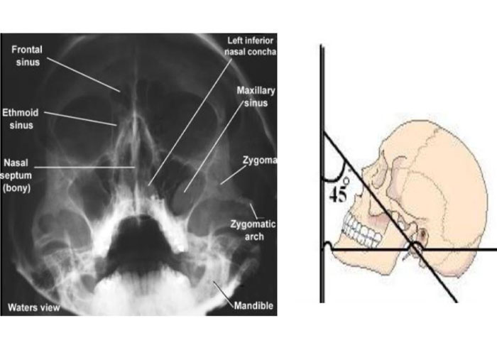
- The image receptor is placed in front of the patient and perpendicular to the midsagittal plane.
- The patient’s head is tilted upward so that the canthomeatal line forms a 45-degree angle with the image receptor.
- If the patient’s mouth is open, the sphenoid sinus will be seen superimposed over the palate.
Position of the Central X-Ray Beam
- The central beam is perpendicular to the image receptor and centered in the area of the maxillary sinuses.

Resultant Image
- The midsagittal plane (represented by an imaginary line extending from the interproximal space of the maxillary central incisors through the nasal septum and the middle of the bridge of the nose) should divide the skull image in two symmetric halves.
- The petrous ridge of the temporal bone should be projected below the floor of the maxillary sinus.
Uses
It can be used to assess for facial fractures, as well as for acute sinusitis. In general, radiographs of the skull and facial bones are rapidly becoming obsolete, being replaced by much more sensitive CT scans.
| Another variation of the waters places the orbitomeatal line at a 37° angle to the image receptor. It is named after the American radiologist Charles Alexander Waters. |
Structures observed
Waters’ view can be used to best visualise a number of structures in the skull.
- Maxillary sinuses.
- Frontal sinuses, seen with an oblique view.
- Ethmoidal cells.
- Sphenoid sinus, seen through the open mouth.
- Odontoid process, where if it is just below the mentum, it confirms adequate extension of the head
Related posts
April 10, 2025
April 9, 2025
April 4, 2025


