- NEED HELP? CALL US NOW
- +919995411505
- [email protected]
Tooth Development
DEVELOPMENTAL STAGES
- enamel organ: growth of dental lamina, 10 in maxilla and 10 in mandible arranged in the form of horse shoe shape
- Dental papilla : ectomesenchymal condensation below the enamel organ
- Dental sac or dental follicle: ectomesenchyme surrounding enamel organ and dental papilla
FATE OF DENTAL LAMINA
- Soon after tooth development the dental lamina starts degenerate
- In third molar region:5 years
- As the tooth develops the connection breaks and islands of epithelial cell remain within the jaws and gingiva – “cell rests of serres”
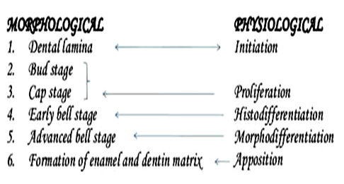
BUD STAGE/ PROLIFERATION
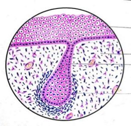
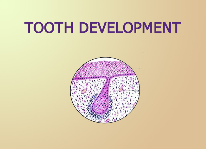
- enamel organ resembles a small bud
- peripherally located low columnar cells and centrally located polygonal cells
- The surrounding mesenchymal cells proliferate results in condensation of cells in 2 areas
– Dental papilla
– Dental sac
CAP STAGE/ PROLIFERATION
- unequal growth in different parts of the tooth bud
- Shallow invagination on the deep surface of bud
- Peripheral cells :cuboidal, cover the convexity of the cap-outer enamel epithelium
- cells in the concavity of the cap:columnar cells-inner enamel epithelium
- Polygonal cells begin to separate due to water being drawn into the enamel organ from the surrounding dental papilla: star shaped: stellate reticulum
- The cell in the center of the enamel organ are densely packed and form the enamel knot: acts as a signaling centers
- a verticle extension of the enamel knot:enamel cord: reservoir of the dividing cell
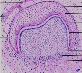
BELL STAGE/ HISTODIFFERENTIATION
- Due to continued uneven growth of the enamel organ it acquires a bell shape
- Shape of the crown is decided
- Inner enamel epithelium:tall columnar cells called ameloblasts
- These cells attached to one another by junctional complexes laterally and to cells in stratum intermedium by desmosomes
- a strong influence on the underlying mesenchymeal cells of dental papilla which later differentiates into odontoblasts
- Stratumintermedium: A few layers of squamous cells, between the inner enamel epithelium and the stellate reticulum
- closely attached by desmosomes & gap junctions
- essential to enamel formation
- Stellate reticulum: expands further due to continued accumulation of intra cellular fluid
- As the enamel formation starts, the stellate reticulum collapses to a narrow zone thereby reducing the distance between the outer and inner enamel epithelium
- Outerenamel epithelium: Low cuboidal cells- These cuboidal cells later assumes a columnar form and produce dentin
- Thrown into folds which are rich in capillary network, this provides a source of nutrition for the enamel organ
- Before the inner enamel epithelium begins to produce enamel. Peripheral cells of the dental papilla differentiate into odontoblasts
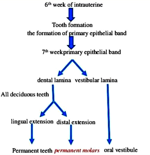
- Dental lamina: extend lingually- successional dental lamina as it give rise to enamel organs of permanent teeth
- Dental sac: Circular arrangement of fibers and resembles a capsule around the enamel organ- form the periodontal ligament fibers that span between the root and the bone
ADVANCEDBELL STAGE/ MORPHODIFFERENTIATION
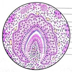
- mineralization and root formation
- The junction between the inner enamel epithelium and odontoblasts outlines the future dentino-enamel junction
- Formation of dentin occurs first along the future dentinoenamel junction in the region of future cusps and proceed pulpally and apically
- After the first layer of dentin is formed, the ameloblasts lay down enamel over the dentin in the future incisal and cuspal areas
- The cervical portion of enamel organ gives rise to hertwig epithelial root sheath (HERS): outlines the future root & thus responsible for the size shape and length & number of roots
- Root formation: begin after enamel & dentin formation has reached the future cementoenamel junction
- HERS remnants: rests of mallasez
MCQS
1. The successors of deciduous teeth develops form_______?
A. Successional lamina
B. Dental lamina
C. Stellate reticulum
D. Neutral ectodermal cells
Answer:A
Related posts
April 10, 2025
April 9, 2025
April 4, 2025


