- NEED HELP? CALL US NOW
- +919995411505
- [email protected]
THYMUS
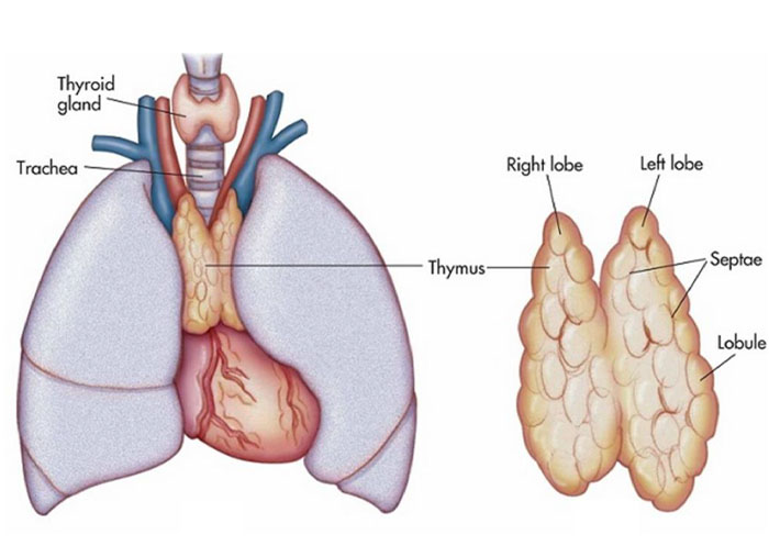
Normal structure
- The thymus gland is a complex lymphoreticular organ lying buried within the mediastinum.
- At birth, the gland weighs 10-35 gm and grows in size upto puberty, following which there is progressive involution in the elderly.
- In the adult, thymus weighs 5-10 gm.
- The gland consists of right and left encapsulated lobes, joined together by fibrous connective tissue.
- Main function of the thymus is in the cell-mediate immunity by T-cells and by secretion of thymic hormones such as thymopoietin and thymosinα1.
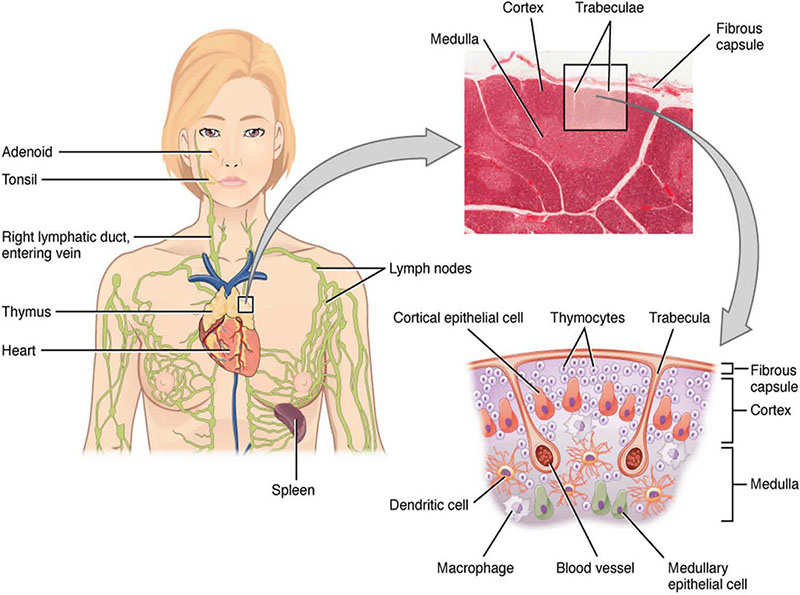
Histologic features
The histologic structure of the lobule shows outer cortex and inner medulla.
Both cortex and medulla contain two types of cells: epithelial cells and
lymphocytes (thymocytes).
Epithelial cells
- Similar throughout the thymus gland.
- Cells have elongated cytoplasmic processes forming network in which
thymocytes and macrophages are found hassall’s corpuscles - Distinctive structures within the medulla composed of onion skin-like
concentrically arranged epithelial cells having central area of
keratinisation.
Thymocytes
- Predominantly present in the cortex.
- Cells include immature t lymphocytes in the cortex and mature t
lymphocytes in the medulla.
THYMIC HYPOPLASIA AND AGENESIS
- Thymic hypoplasia and agenesis are acquired and congenital disorders respectively in which the gland is either unusually small or absent.
- These conditions are various types of hereditary (primary) immunodeficiency diseases such as DiGeorge’s syndrome, severe combined immunodeficiency and reticular dysgenesis.
- Acquired hypoplasia occurs as an ageing phenomenon or may occur in the young due to severe stress, malnutrition, irradiation, therapy with cytotoxic drugs and glucocorticoids.
THYMIC HYPERPLASIA
- Enlargement of the thymus or failure to involute
- appearance of lymphoid follicles in the medulla of the thymus and is called thymic follicular hyperplasia.
- Most common cause of follicular hyperplasia of the thymus is myasthenia gravis.
- Less common causes are: Addison’s disease, Graves’ disease, rheumatoid arthritis, SLE, scleroderma and cirrhosis of liver.
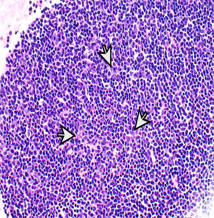
THYMOMA
- Most common primary tumour present in the anterosuperior mediastinum is thymoma.
- Although thymus is a lymphoepithelial organ, the term thymoma is used for the tumour of epithelial origin.
- Most of the patients are adults.
- In about half the cases, thymoma remains asymptomatic and is accidentally discovered in X-rays.
- Other patients have associated conditions like myasthenia gravis or local symptoms such as cough, dyspnoea and chest pain.
- Thymomas are known for their association with paraneoplastic syndrome
Morphologic features.
Grossly, the tumour is spherical, measuring 5-10 cm in diameter with an average weight of 150 gm.
Sectioned surface is soft, yellowish, lobulated and may be either homogeneous or contain cysts
Microscopically, the tumour has a thick fibrous capsule from which extend collagenous septa into the tumour dividing it into lobules.
The tumour consists of neoplastic epithelial cells and variable number of nonneoplastic lymphocytes.
Types
- Benign thymoma is more common.
- Malignant thymoma is less common and is further of 2 types:
| Type 1 is cytologically benign looking but aggressive and invades the mediastinal structures locally | Type 2 is also called thymic carcinoma and has cytologic features of cancer |