- NEED HELP? CALL US NOW
- +919995411505
- [email protected]
Temporomandibular Joint
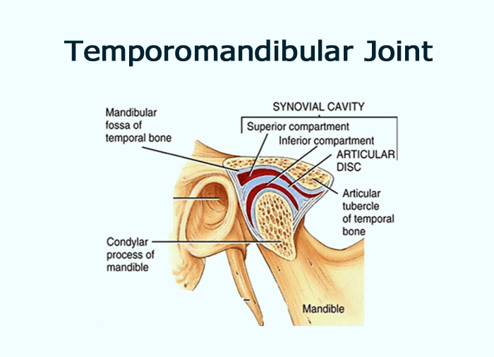
| Compartments | Superior (translational movement) and inferior compartments (rotational movement) |
| Joint capsule | Limits/borders - border of mandibular fossa and neck of the mandible above the pterygoid fovea |
| Ligaments | Collateral, temporomandibular, stylomandibular, and sphenomandibular ligaments |
| Vascular supply | Deep auricular, superficial temporal, and anterior tympanic arteries |
| Innervation | Mandibular, masseteric, and deep temporal nerves, together with the otic and superior cervical ganglions |
Compartments
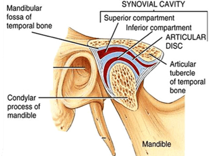
The joint is separated into a superior and an inferior compartment by the articular disc.- The superior compartment is bordered superiorly by the mandibular fossa of the temporal boneand inferiorly by the articular disc itself. It contains 1.2 mL of synovial fluid and is responsible for the translational movement of the joint.
- The inferior compartment has the articular disc as a superior border and the condyle of the mandibleas an inferior border. It is slightly smaller with an average synovial fluid volume of 0.9 mL and allows rotational movements.
Capsule and ligaments
The joint capsule originates from the border of the mandibular fossa, encloses the articular tubercle of temporal bone and inserts at the neck of mandible above the pterygoid fovea. It is so loose that the mandible can naturally dislocate anteriorly without damaging any fibres of the capsule.
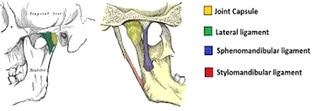
The TMJ is supported by the following ligaments:
- The medial and lateral collateral ligaments(also known as the discal ligaments) help connect the medial and lateral sides of the articular disc to the same side of the condyle.
- The temporomandibular ligamentis located on the lateral aspect of the capsule and its function includes preventing the lateral or posterior displacement of the condyle.
- The stylomandibular ligamentarises from the styloid process and attaches to the mandibular angle. It is responsible for allowing the mandible to protrude.
- The sphenomandibular ligamentstretches between the spine of the sphenoid bone and the lingua of the mandible. It contributes to the limitation of extensive protrusive movements and jaw opening.
Vascular supply
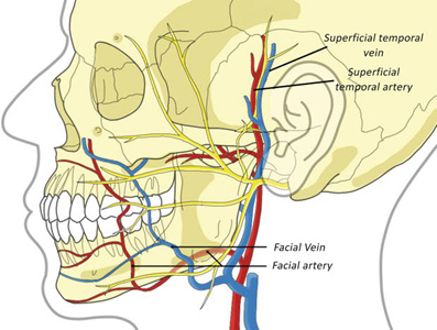
The TMJ is supplied by three arteries. The main supply comes from the deep auricular artery (from the maxillary artery) and the superficial temporal artery (a terminal branch of the external carotid artery). In addition the joint is provided by the anterior tympanic artery (also a branch of the maxillary artery). The venous blood drains through the superficial temporal vein and the maxillary vein.
Innervation
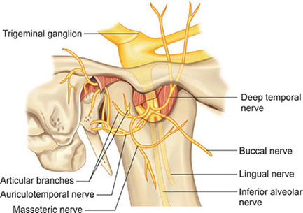
The mandibular nerve (third branch of the trigeminal nerve) provides the main nerve supply of the TMJ. Additional innervation comes from the masseteric nerve and deep temporal nerves. Parasympathetic fibres of the optic ganglion stimulate the synovial production. Sympathetic neurons from the superior cervical ganglion reach the joint along the vessels and play a role in pain reception and the monitoring of the blood volume.
MCQs
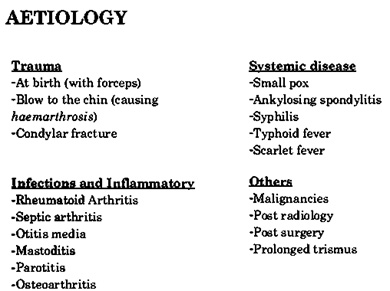
1. Common cause of TMJ ankyloses isa) Trauma
b) Development disturbances
c) Infections
d) Atrophy
Answer: A
Related posts
April 10, 2025
April 9, 2025
April 4, 2025


