- NEED HELP? CALL US NOW
- +919995411505
- [email protected]
SUBMENTOVERTEX (BASE) PROJECTION

Image Receptor and Patient Placement
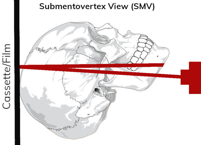
- The image receptor is positioned parallel to patient’s transverse plane and
perpendicular to the midsagittal and coronal planes. - To achieve this, the patient’s neck is extended as far backward as
possible, with the canthomeatal line forming a 10-degree angle with the
image receptor.
Position of the Central X-Ray Beam
- The central beam is perpendicular to the image receptor, directed from
below the mandible toward the vertex of the skull (hence the name
submentovertex, or SMV ), - Centered about 2 cm anterior to a line connecting the right and left
condyles.

Resultant Image
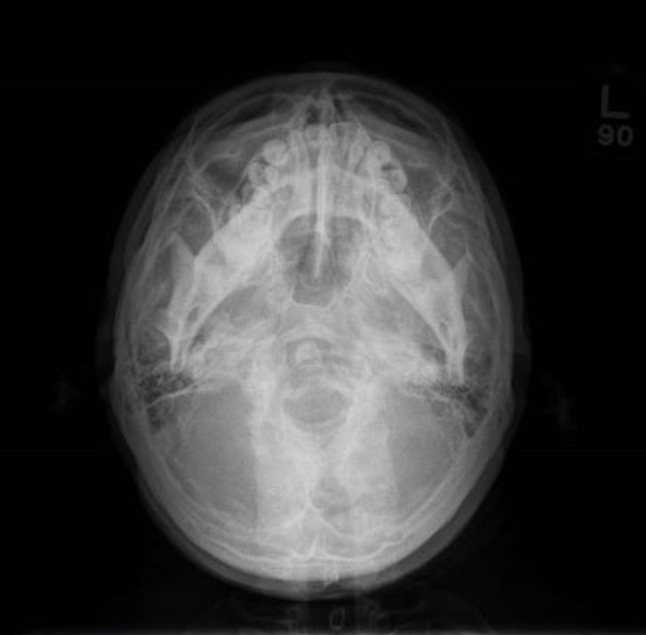
- The midsagittal plane (represented by an imaginary line extending from the interproximal space of the maxillary central incisors through the nasal septum, to the middle of the anterior arch of the atlas, and to the dens) should divide the skull image in two symmetric halves.
- The buccal and lingual cortical plates of the mandible should be projected as uniform opaque lines.
- An underexposed view is required for the evaluation of the zygomatic arches because they will be overexposed or “ burned out ” on radiographs obtained with normal exposure factors

Clinical view
This view is useful in assessing potential pathology from trauma or disease progression to the basal skull structures, including the foramen ovale, foramen spinosum and sphenoid sinuses.
It is imperative that any cervical spine subluxations or fractures on acute trauma patients are excluded before proceeding with this view.
Related posts
April 10, 2025
April 9, 2025
April 4, 2025


