- NEED HELP? CALL US NOW
- +919995411505
- [email protected]

- PET is a more advanced imaging modality in nuclear medicine.
- PET, which is reported to have a sensitivity nearly 100 times that of a gamma camera, relies on positron-emitting radionuclides generated in a cyclotron.
- The utility of PET is based not only on its sensitivity but also on the fact that the most commonly used radionuclides ( 11 C, 13 N, 15 O, 18 F) are isotopes of elements that occur naturally in organic molecules.
- Although fluorine does not technically fit into this category, it is a chemical substitute for hydrogen.
Mechanism
- These radionuclides are used as is, or more commonly, incorporated into a radiopharmaceutical such as glucose or amino acids by use of a medical cyclotron.
- After the radiopharmaceutical is injected into the patient, the isotope distributes within the body ’s tissue according to the carrier molecule and emits a positron.
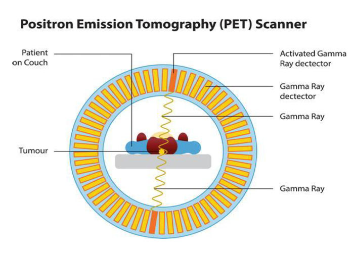
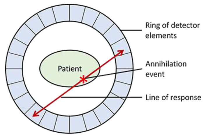
- This positron then interacts with a free electron and mutual annihilation occurs, resulting in the production of two 551- keV photons emitted at 180 degrees to each other.

Parts
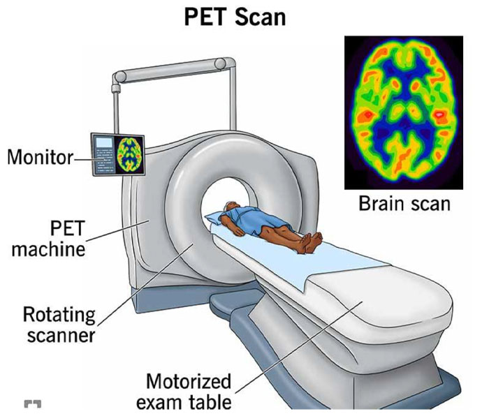
- The PET scanner consists of a ring of many detectors in a circle around the patient.
- The detector crystals are often made of bismuth germinate.
- Electronically coupled opposing detectors simultaneously identify the pair of γ photons using coincidence detection circuits that measure events within 10 to 20 nanoseconds.
- The annihilation event is thus known to have occurred along the line joining the two detectors.

Raw PET scan data consist of a number of these coincidence lines, which are reorganized into projections that identify where isotope is concentrated within the patient.
The spatial resolution of a PET scanner is about 5 mm. PET is useful in skeletal imaging for assessing primary bone tumors, locating metastases in bone, and detecting osteomyelitis.

For instance, 18 F-fl uoro-2-deoxyglucose ( 18 F-FDG) is a radiopharmaceutical commonly used for studying glucose use in the brain and heart and to look for cancer metastases.
PET images are often fused with CT scans to facilitate anatomic localization of radionuclide.
The PET/CT combination has been shown to be quite helpful in staging and treatment planning of squamous cell carcinoma in the head and neck


