- NEED HELP? CALL US NOW
- +919995411505
- [email protected]
Patent ductus arteriosus (PDA)

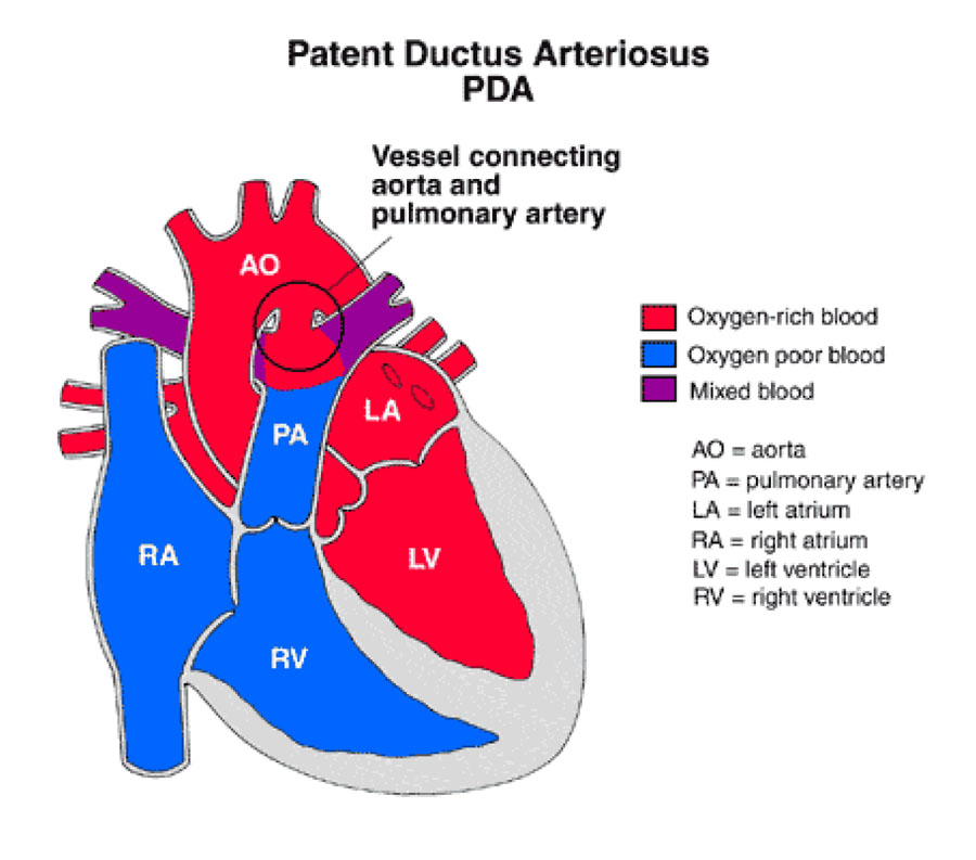
Is a persistent opening between the two major blood vessels leading from the heart. The heart problem is present from birth. That means it is a congenital heart defect.
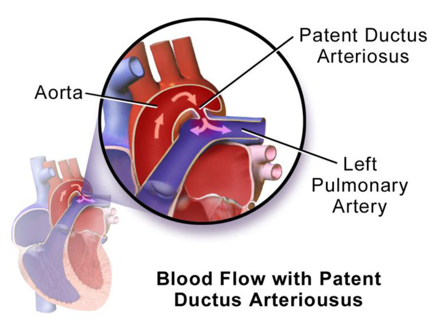
An opening called the ductus arteriosus is part of a baby's blood flow system in the womb. It usually closes shortly after birth. If it remains open, it's called a patent ductus arteriosus.

- Before a baby is born, the fetus's blood does not need to go to the lungs to get oxygenated.
- The ductus arteriosus is a hole that allows the blood to skip the circulation to the lungs.
- However, when the baby is born, the blood must receive oxygen in the lungs and this hole is supposed to close.
- If the ductus arteriosus is still open (or patent) the blood may skip this necessary step of circulation.
- The open hole is called the patent ductus arteriosus.
- Normally the heart's left side only pumps blood to the body, and the right side only pumps blood to the lungs.
- In a child with PDA, extra blood gets pumped from the body artery (aorta) into the lung (pulmonary) arteries.
- If the PDA is large, the extra blood being pumped into the lung arteries makes the heart and lungs work harder and the lungs can become congested.

CLINICAL FEATURES
- The history of the mother’s pregnancy and of perinatal events may provide clues associated with a high incidence of PDA, such as exposure to rubella in the first trimester in a non-immunized mother.
- PDA is also more common in premature infants, especially those with birth asphyxia or respiratory distress.
- Symptoms depends on the degree of left-to- right shunting; and it is determined by the size of the PDA, ductal resistance, cardiac output, as well as the systemic and pulmonary vascular resistances.
- Patients with small PDA are asymptomatic.
- With larger PDAs, symptoms may develop.
- The most common symptom is exercise intolerance followed by dyspnea, peripheral edema, and palpitations.
SIGNS
- Tachycardia and tachypnea may be present in infants with CHF.
- Bounding peripheral pulses.
- Wide pulse pressure.
- Hyperactive precordium: With a large shunt.
- Systolic thrill: may be present at the upper left sternal border.
- The P2 is usually normal, but its intensity may be accentuated if pulmonary hypertension is present.
INVESTIGATION
- With small shunt the ECG is normal.
- Left ventricular hypertrophy of the volume overload type, with deep Q waves and increased R- wave voltage in the left precordial leads, is noted with increasing shunt size with left ventricular volume overload.
- Right ventricular hypertrophy is seen with pulmonary hypertension.
- Chest radiographs may be normal with a small-shunt PDA.
- Cardiomegaly of varying degrees occurs in moderate- to large-shunt PDA with enlargement of the LA, LV, and ascending aorta.
- Pulmonary vascular markings are increased.
- With pulmonary vascular obstructive disease, the heart size becomes normal, with a marked prominence of the PA segment and hilar vessels.
- TTE has a 42% sensitivity and 100% specificity for the diagnosis of PDA.
- On 2-D echo, the left-sided chambers (LA and LV) are dilated due to increased venous return from the pulmonary circulation.
- This constitutes left ventricular volume overload.
- Due to dilatation of left atrium, the ratio between size of the left atrium and proximal aorta (LA : Ao ratio) exceeds 1.3.
- Continuous wave Doppler echocardiogram positioned through the PDA, showing retrograde flow throughout the cardiac cycle Pulsed-Doppler echocardiogram shows increased diastolic flow in the branch pulmonary artery
- Catheter may easily pass from PA to Ao through the PDA.
- It gives a specific appearance “Hair pin” appearance.
- Steps up of O2 saturation in PA in comparison to RA.
- Pressure study: RV & PA pressure is normal, but elevated in large PDA.
- PVR is normal in infant & children but elevated in adult.
- LV graphy: to see associated VSD
- Aortography: to see PDA & associated CoA
- Magnetic resonance imaging (MRI) and computed tomography may be useful in defining the anatomy in patients with unusual PDA geometry and in patients with associated abnormalities of the aortic arch.
| ECG | |
| CXR | |
| Echocardiogram | |
| Cardiac Catheterization Catheter trajectory |
|
| Oxymetry | |
| MRI & CT |
MANAGEMENT
If the PDA isn't causing problems, a catheter treatment to close the opening may be done when the baby is older. Open-heart surgery to close the PDA . This treatment is called surgical closure. Heart surgery may be needed if medicine doesn't work or the PDA is large or causing complications.
Related posts
April 10, 2025
April 9, 2025
April 4, 2025


