- NEED HELP? CALL US NOW
- +919995411505
- [email protected]
Odontogenic Keratocyst

DEFINITION
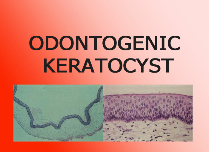
According to WHO it is a benign uni or multicystic intraosseous tumor of odontogenic origin (dental lamina and its remnant) with characteristic lining of parakeratinised stratified squamous epithelium & potential for aggressive and infiltrative behavior.
CAUSES
- There is general agreement that OKCs develop from dental lamina remnants in the mandible and maxilla. However, an origin of this cyst from extension of basal cells of the overlying oral epithelium has also been suggested.
- Genetic
HISTOPATHOLOGY

- The epithelial lining is uniformly thin, generally ranging from 8 to 10 cell layers thick.
- The basal layer exhibits a characteristic palisaded pattern with polarized and intensely stained nuclei of uniform diameter. The luminal epithelial cells are parakeratinized and produce an uneven or corrugated
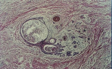
- budding of the basal cells into the C.T wall and microcyst
- The fibrous connective tissue component of the cyst wall is often free of inflammatory cell infiltrate and is relatively thin.

CLINICAL FEATURES
- Age: Any age , especially adults.
- Location: Mandibular molar, ramus area favored; may be found dentigerous, in position of lateral root, periapical, or primordial cyst.
- OKCs are relatively common jaw cysts. They occur at any age and have a peak incidence within the second and third decades.
RADIOGRAPHIC FEATURES
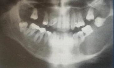
- Location: The most common is the posterior body of the mandible (90% posterior to the canines) and ramus (more than 50%). This type of cyst occasionally has the same pericoronal position as dentigerous cyst.
- Periphery and shape: Usually with a cortical border unless becomes secondarily infected. The cyst may have a smooth (round or oval shape), or it may have a scalloped outline.
- Internal structure:
- Most commonly is radiolucent.
- The cystic cavity contain keratin.
- In some cases curved internal septa may be present, giving the lesion a multilocular appearance.
- The effects on surrounding structures: It grows along the internal aspect of the jaws, causing minimal expansion except for the upper ramus and coronoid process, where considerable expansion may occur. OKCs can displace and resorb teeth but to a slightly lesser degree than dentigerous cysts. The inferior alveolar nerve canal may be displaced inferiorly. In the maxilla this cyst can invaginate and occupy the entire maxillary antrum.

DIFFERENTIAL DIAGNOSIS
- Histologically :
- Myxoma
- Ameloblastoma
- central giant cell granuloma
- odontogenic cysts.
- Radiographically :
- Dentigerous cyst
- Residual cysts, redicular cyst
- Lateral periodontal cysts
- Primordial cyst
- Globulomaxillary cyst
- Unicystic ameloblastoma
- A-V malformation
- Fibroosseous lesion at initial stages
SYNDROMES ASSOCIATED WITH MULTIPLE OKC
- Nevoid Basal cell carcinoma syndrome (NBCCS)
- Gorlingoltz syndrome
- Marfans syndrome
- Ehlers danlosdanlos syndrome
- Noonans syndrome
- Orofacial digital syndrome
- Simpsongolabi-behmel syndrome
TREATMENT
Depend upon patient age, size and location of cyst, soft tissue involvement and histological variant of lesion
CLASSIFICATION OF TREATMENT PLAN
- Enucleation
- Enucleation with carnoy’ solution
- Enucleation with perepheral ostectomy
- Enucleation with carnoy’ solution and perepheral ostectomy
- Enucleation & cryotherapy
- Marsupialization
- Marsupialization with cystectomy
- Resection
- Enucleation: This technique involves complete removal of the cystic sac and healing of the wound by primary intention. This is the most satisfactory method of treatment of a cyst and is indicated in all cases where cysts are involved, whose wall may be removed without damaging adjacent teeth and other anatomic structures.
- Marsupialization: This method is usually employed for the removal of large cysts and entails opening a surgical window at an appropriate site above the lesion. In order to create the surgical window, initially a circular incision is made, which includes the mucoperiosteum, the underlying perforated (usually) bone, and the respective wall of the cystic sac.
MCQs
- Multiple odontogenic keratocyst are associated with:____?
- Gardner’s syndrome
- Gorlin-Goltz syndrome
- Goldenhar’s syndrome
- Grinspan syndrome
Answer: B
Related posts
April 10, 2025
April 9, 2025
April 4, 2025


