- NEED HELP? CALL US NOW
- +919995411505
- [email protected]

A radiographic review
Synonym
Vitamin D – resistant rickets and hypophosphatemic rickets
- Vitamin D-resistant hypophosphatemic rickets, also known as familial or hereditary hypophosphatemic rickets (HHR).
- Produce renal tubular disorders resulting in excessive loss of phosphorus.
- There is a failure to reabsorb phosphorus in the distal renal tubules, resulting in a decrease in serum phosphorus (hypophosphatemia).
- Two causes need to be considered in renal phosphate wasting: on the one hand caused by a primary renal tubular defect.
- On the other hand as a secondary effect due to increased fibroblast growth factor 23 (FGF23) signaling, as is seen in HHR.
- The increased FGF-23 has a dual effect in HHR. Firstly, it inhibits renal phosphate resorption in the proximal tubule of the nephron by decreasing the number of sodium-transporters.
- Secondly FGF-23 also inhibits 1-a-hydroxylase, which diminishes vitamin D activation.
- Consequently, renal phosphate reabsorption and intestinal uptake of phosphate and calcium will decrease respectively.
- Normal calcification of the osseous structures requires the correct amount and ratio of serum calcium and phosphorus.
- Multiple myeloma may induce hypophosphatemia as a result of secondary damage to the kidneys.
Clinical Features
- Children with hypophosphatemia show reduced growth and rickets like bony changes.
- These include bowing of the legs, enlarged epiphyses, and skull changes. Adults have bone pain, muscle weakness, and vertebral fractures.
Radiographic Features
General Radiographic Features.
- In children with hypophosphatemia, radiographic findings are indistinguishable from those of rickets. In adults the long bones may show persistent deformity, fractures, or pseudofractures.
Radiographic Features of the Jaws.
- The jaws are usually osteoporotic and in extreme cases are remarkably radiolucent. Cortical boundaries may be unusually radiolucent or not apparent .
- Fewer visible trabeculae and a granular trabecular pattern.
Radiographic Features Associated with the Teeth.
- The teeth may be poorly formed, with thin enamel caps and large pulp chambers and root canals.
- Periapical and periodontal abscesses occur frequently.
- Occurrence of periapical rarefying osteitis without an etiology may be a result of large pulp chambers and defects in the formation of dentin.
- If the disease is severe, the patient has premature loss of the teeth.
- The lamina dura may become sparse, and cortical boundaries around tooth crypts may be thin or entirely absent.
Few case report radiographic pictures
CASE 1
Clinical and radiographic dental manifestations
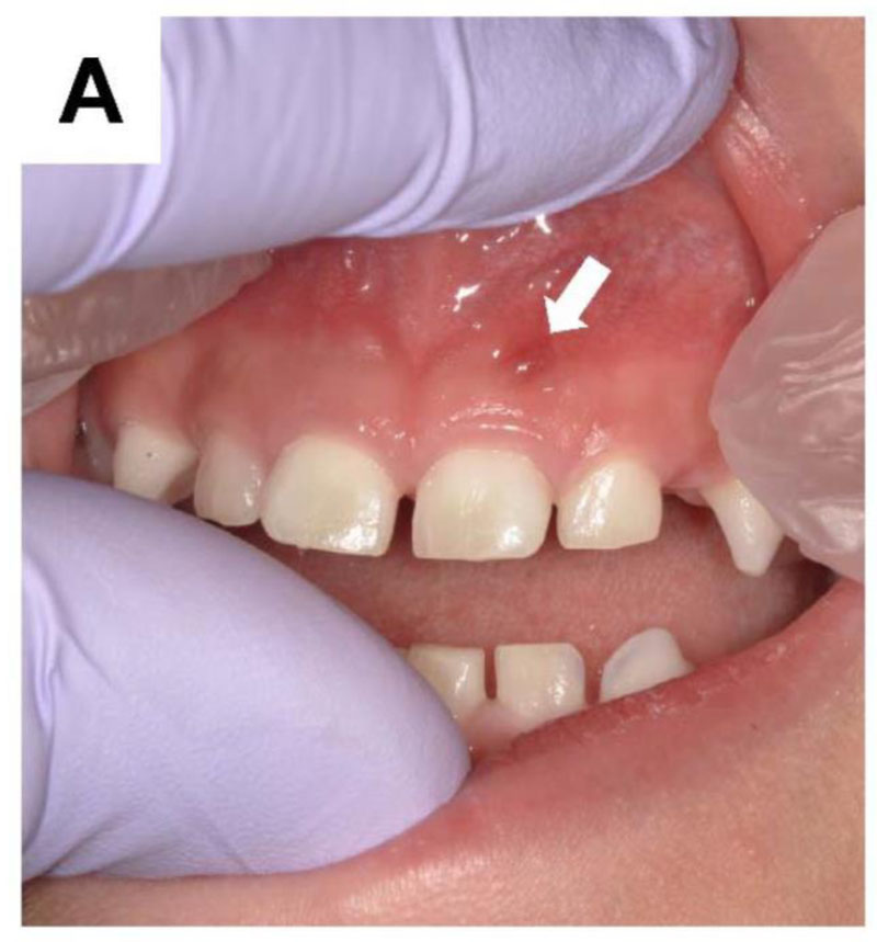
(A) Oral photograph of male patient aged 4 years 4 months showing a periapical gingival abscess (white arrow) corresponding to the primary maxillary left central incisor.
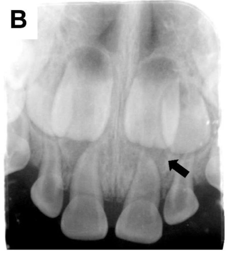
(B) Periapical radiograph showing radiolucency (black arrow) around the periapical region of the primary maxillary left central incisor.
CASE 2
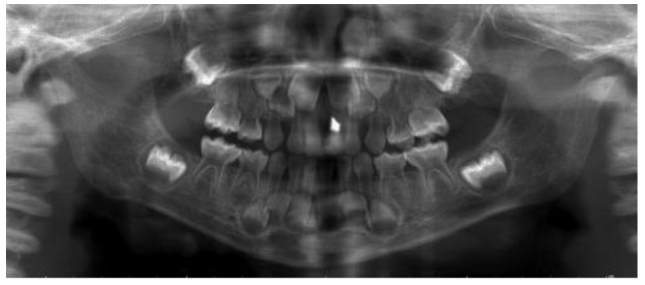
A 1Y5M boy with spontaneous pain in the maxillary anterior region. Clinical examinations led to diagnosis of acute periapical periodontitis in the maxillary left primary central incisor, for which root canal treatment was performed. Thereafter, a pediatrician diagnosed XLH based on elevated ALP in a blood test at 1Y10M.
CASE 3
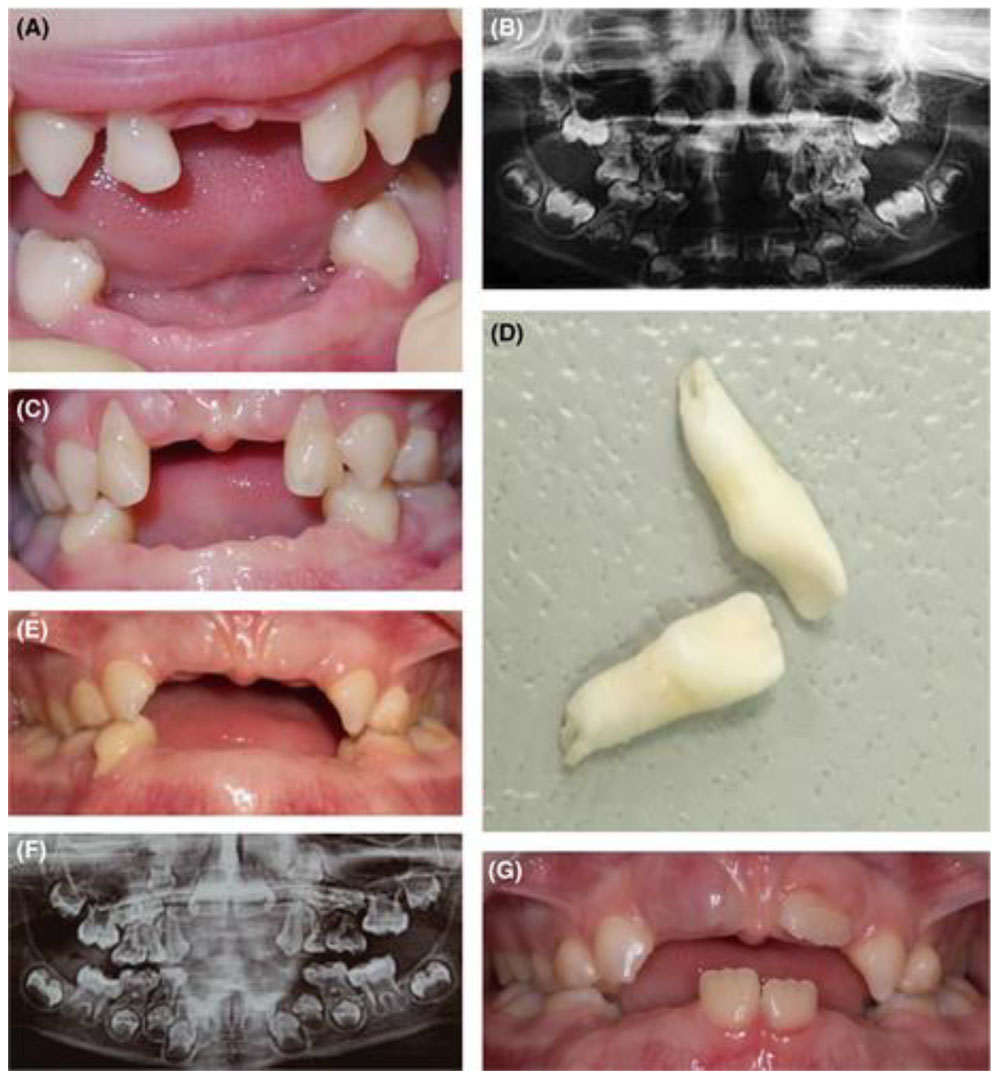
The mother complained that many of the primary teeth of her daughter had spontaneously fallen out. At the age of 11 months, the lower central incisors erupted, and at 12 months, they were shed. At the age of 20 months, the lower lateral incisors fell out. By the age of 2 years, the upper central incisors had fallen out. At 3 years and 6 months, the lower canines were shed. Nevertheless, the mother reported no history of trauma.
Physical examination revealed short stature, a bulging frontal bone, lower limb bow, and. Intraoral examination revealed an upper arch with the absence of primary central incisors and caries-free primary dentition.
The primary lateral upper incisors were Grade 2 mobile. There was minimal gingival inflammation. The lower arch exhibited the absence of primary frontal teeth due to premature shedding. The upper lateral incisors and the first lower left molar were lost spontaneously because of extreme mobility despite the start of enzyme replacement therapy.
Radiographically, enlarged pulp chambers and shape abnormalities of the permanent teeth crowns were revealed. Horizontal alveolar bone loss reached nearly half of the root length.
