- NEED HELP? CALL US NOW
- +919995411505
- [email protected]

Hertwig’s Epithelial Root Sheath (HERS) was first discovered as a bi-layered cell sheath surrounding the roots of amphibian teeth (e.g. in Triturus and Salamandra)(Hertwig, 1874).
HERS descends from the oral epithelium and the enamel organ to form a collar around the cervical part of the amphibian tooth root
The Hertwig epithelial root sheath (HERS) or epithelial root sheath is a proliferation of epithelial cells located at the cervical loop of the enamel organ in a developing tooth.
Hertwig epithelial root sheath initiates the formation of dentin in the root of a tooth by causing the differentiation of odontoblasts from the dental papilla.
STRUCTURE
- Consists of only inner and outer enamel epithelium
- Before root formation begins epithelial diaphragm forms

FUNCTION
- The Hertwig’s epithelial root sheath is a transient structure formed in the early period of root formation.
- It has an important role in regulation and maintenance of the periodontal ligament space,
- Prevention of root resorption and ankylosis,
- Maintenance of periodontal ligament homeostasis,
- Induction of acellular cementum formation,
- As a stem cell in periodontal regeneration and also as a stimulant in periodontal regeneration
FATE OF HERS
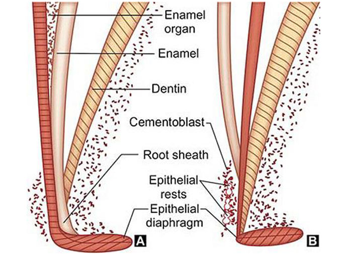
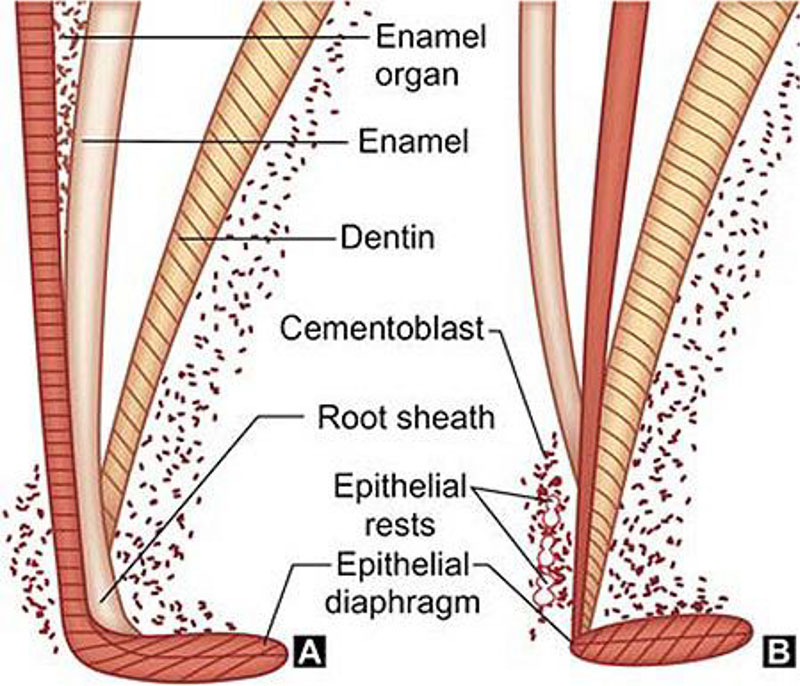
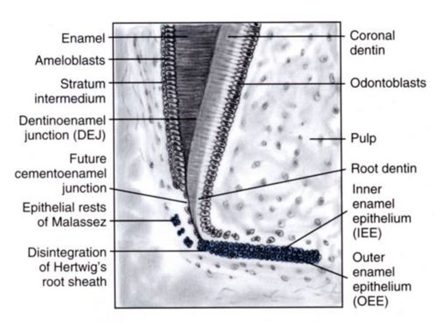
The root sheath eventually disintegrates with the periodontal ligament, but residual pieces that do not completely disappear are seen as epithelial cell rests of Malassez (ERM).These rests can become cystic, presenting future periodontal infections

ROOT SHEATH DEVELOPMENT
- The development of the Hertwig’s epithelial root sheath originates at the end of the crown stage in tooth development
- The ectodermal derivatives, inner and outer enamel epithelium of the enamel organ (devoid of stratum intermedium and stellate reticulum) proliferate and give rise to a double-layered, tubelike sleeve of epithelial cells, known as the Hertwig’s epithelial root sheath
- Epithelial cells of the inner and outer enamel epithelium proliferate from the cervical loop to form two layers of epithelium called Hertwig’s root sheath
- The first formed part of the root sheath bends to form a disc like structure
- The rim of this disc like structure is called the epithelial diaphragm
- The epithelial diaphragm encloses the primary apical foramen
- After the formation of epithelial root sheath and the epithelial diaphragm the root grows in length
- The diaphragm maintains a constant size while the root sheath grows in length at the angle of the diaphragm and not at its tip
- The cells of the lengthening root sheath induce the adjacent dental papilla cells to differentiate into odontoblasts
- The newly formed odontoblasts then form the root dentine
- As the root lengthens the crown moves occlusally
DEVELOPMENTAL ANOMALIES/PATHOLOGIES ASSOCIATED WITH HERS
- Enamel pearls: Some cells of HERS remain adherent to the dentin surface and differentiate into fully functioning ameloblasts, producing enamel. Such droplets of enamel, called ENAMEL PEARLS,are sometimes found in the area of furcation of roots of permanent molars
- Periapical cyst: The remnants of HERS in the periapical region have the potential for reactive proliferation in response to an adjacent focus of infection resulting in formation of Periapical cyst
- Accessory root canals: If the continuity of HERS is broken prior to dentin formation,a defect in the dentin wall of the pulp consequences. Such defects are found in the furcation or on any point of the root itself resulting in development of accessory root canals opening on periodontal surface of the root
- Periodontal pocket: The periodontal pocket is a focus of infection which occurs as a localized area of chronic inflammation in the connective tissues adjacent to the epithelial rests


