- NEED HELP? CALL US NOW
- +919995411505
- [email protected]
Frequently asked NEET Questions

1. Sensory supply to nasal cavity is by
- Trigeminal
- Facial
- Frontal
- Ethmoid
ANSWER. a
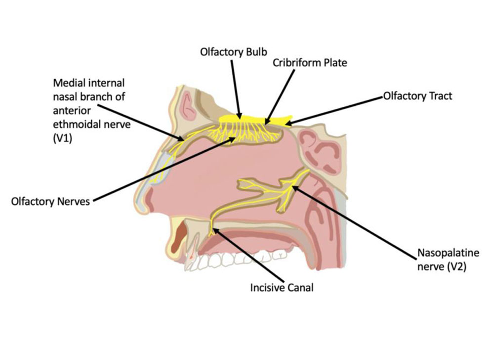
- The innervation of the nose can be functionally divided into special and general innervation.
- Special sensory innervation refers to the ability of the nose to smell.
- This is carried out by the olfactory nerves.
- General sensation is carried by the trigeminal nerve (CN V)
- Serous glands in the nasal mucosa which produce fluid that constantly lubricates the nose walls are innervated by the parasympathetic fibers of the facial nerve (CN VII). Sympathetic innervation comes from T1 level of spinal cord and is intended for regulation of blood flow through mucosa.


2. Danger space is
- Carotid sheath
- Space between the alar plate and prevertebral fascia
- Posterior to prevertebral area
- Space posterior to carotid sheath in posterior triangle
- The cervical fascia can be divided anatomically into superficial and deep fascia.
- The superficial fascia consists of skin, subcutaneous tissue and the platysma, and the deep fascia is further divided into superficial, middle and deep layers.
ANSWER. b
THE SUPERFICIAL LAYER (OR INVESTING LAYER)
- Covers the submaxillary and parotid glands as well as muscles deep to the platysma.
- This layer encloses the submandibular and masticator spaces, which can be a focus of dental or submandibular infections.
THE MIDDLE LAYER (OR PRETRACHEAL LAYER)
- encloses the visceral organs of the neck, namely (from anterior to posterior), the thyroid and parathyroid glands, the larynx and trachea, the pharynx, and oesophagus.
THE DEEP LAYER (OR PREVERTEBRAL LAYER)
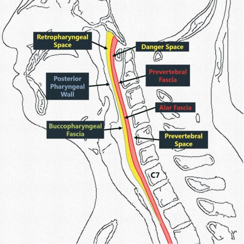
- Covers the vertebral column and the paravertebral muscles There is a space between the middle and deep layers anteriorly, termed the retropharyngeal space, which is subdivided by a thin membrane called the alar fascia.
- The deep cervical fascial layers establish two clinically significant potential spaces: the retropharyngeal and danger spaces
- The danger space lies behind the true retropharyngeal space.
- Boundaries:
- Superiorly: clivus
- Inferiorly: posterior mediastinum at the level of the diaphragm
- Anteriorly: alar fascia
- Posteriorly: prevertebral fascia

3. Function of carotid body is
- Sense O2 partial pressure in arterial blood
- Sense O2 partial pressure in venous blood
- Regulate tissue fluids
- Regulate haemoglobin synthesis
ANSWER. a
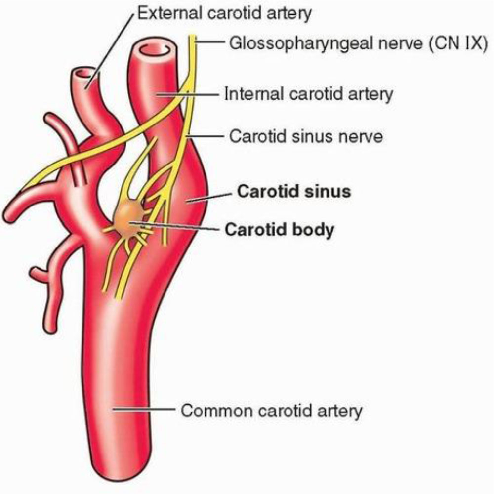
- The carotid bodies are small (≈2 mm diameter in humans) sensory organs located near the carotid sinus at the bifurcation of the common carotid artery at the base of the skull.
- The aortic bodies are on the aortic arch near the aortic arch baroreceptors.
- Carotid body is a peripheral chemoreceptor that monitors arterial blood gas tensions and pH.
- Its main function is to contribute to the regulation of breathing although there is evidence for a reflex influence on the pulmonary circulation and the kidney.
- Carotid body sensory activity increases in response to arterial hypoxemia and the ensuing chemoreflex regulates vital homeostatic functions

4. Factor presents both in serum and plasma
- Prothrombin
- Factor V
- Factor VII
- Factor VIII
ANSWER. c
5. 75% of brain growth is complete by
- 6 months
- 2 year
- 5 year
- 12 year
ANSWER. b
- At birth, the average baby’s brain is about a quarter of the size of the average adult brain.
- It doubles in size in the first year. It keeps growing to about 75% of adult size by age 2 and 90% – nearly full grown – by age 5.
- The brain is the command center of the human body.
- A newborn baby has all of the brain cells (neurons) they’ll have for the rest of their life, but it’s the connections between these cells that really make the brain work. Brain connections enable us to move, think, communicate and do just about everything.
- The early childhood years are crucial for making these connections.
- At least one million new neural connections (synapses) are made every second, more than at any other time in life.
Related posts
April 10, 2025
April 9, 2025
April 4, 2025


