- NEED HELP? CALL US NOW
- +919995411505
- [email protected]

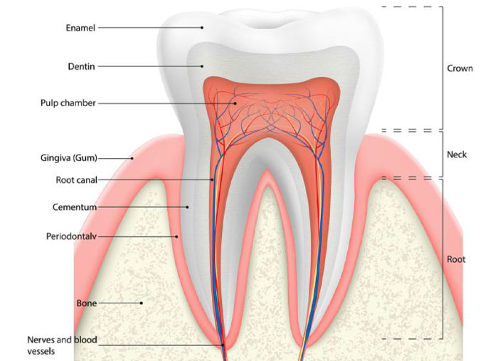
Dentin or dentine is a layer of material that lies immediately underneath the enamel of the tooth. It is one of the four major components of the tooth which comprises:
- The outer hard enamel
- The dentin underneath the enamel
- The dental pulp that lies soft and encased within the dentin
- The cementum that lies over the dentin and helps attach the tooth to the jaw

Dentin is a calcified tissue of the body and, along with enamel, cementum, and pulp, is one of the four major components of teeth
Dentin Physically and chemically, it closely resembles bone
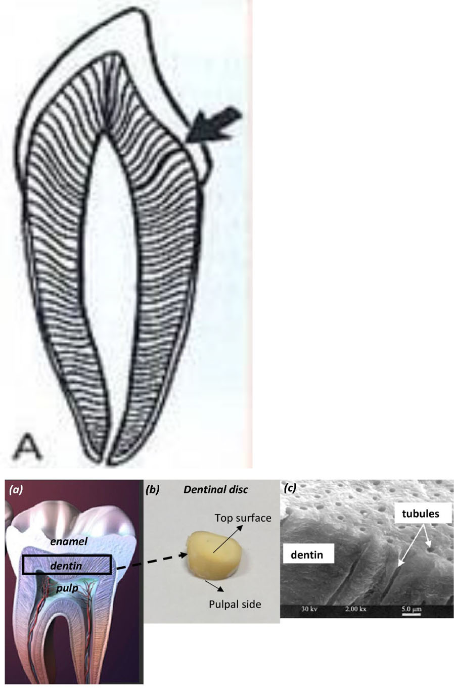
Said to be a living tissue since the tubules present in it contains processes of specialised cells, the odontoblasts.
Main morphologic difference between bone and dentin is that some of the osteoblasts exist on the surface of the bone and when one of the cells becomes enclosed within its matrix, it is called an osteocyte.
Development of dentin
- Prior to enamel formation, dentine formation begins through a process known as dentinogenesis, and this process continues throughout a person's life even after the tooth has fully developed.
- The dentin is formed from the odontoblasts
- The odontoblasts first form predentin which then matures into dentin
Physical properties
Colour
- Young age -light yellow
- With advancing age -becomes darker
Consistency
- Elastic and resilient
- Harder than bone but softer than enamel
Tensile strength : 40mpa
Compressive strength : 266mpa
Composition
- Dentin has 35% organic, 65% inorganic and water by weight .
- The organic matrix of dentin is collagenous
- It provides resiliency to the crown which is necessary to withstand the forces of mastication
- The principle inorganic component of dentin is hydroxyapatite crystals The high mineral content of dentin makes it harder than bone and cementum but softer than enamel .
- The knoop hardness for dentin is approximately 68
Organic substances |
Inorganic substances |
|
|
DENTINAL TUBULES
- The course of the dentinal tubules follow a gentle curve in the crown where it resembles an S shape
- Starts at right angles at the pulpal surface, the first convexity of this doubly curved course is directed towards the apex of the tooth
- These tubules end perpendicular to the DEJ & CDJ
- It is almost straight near the root tip and along the incisal edges and cusp
- Dentin thickness ranges from 3-10mm or more
- Ratio between outer and inner surfaces of dentine is about 5:1
- No. of tubules per square millimeter varies from 15000 at the DEJ to 65000 at the pulp – density and diameter increases with depth
- There are more tubules per unit area in the crown than in the root

Peritubular dentin
- Dentin that immediately surrounds the dentinal tubules
- Found throughout the thickness of dentin except near the pulp
- More highly mineralized (9% more) than intertubular dentin
- Thin organic membrane rich in glycosaminoglycans on the inner side
Intertubular dentin
- Located between the zones of peritubular dentin
- One half of its volume is organic matrix, specifically collagen fibres
- The fibrils range from 0.5-0.2µm in diameter and exhibit crossbanding at 64µm intervals
- HA crystals are formed along the fibres with their long axis oriented parallel to the collagen fibres
- Well mineralised, Provide tensile strength to dentin
Predentin
- 2µm - 6µm wide layer of unmineralized dentin matrix located adjacent to pulp tissue
- First formed dentin
- Thickness depends on the activity of odontoblasts
Odontoblastic processes
- Cytoplasmic extensions of the odontoblasts
- The odontoblasts reside in the peripheral pulp at the pulp-predentin border and their processes extend into the dentinal tubules
- The processes are largest in diameter near the pulp and taper further into dentin
- The odontoblast cell bodies are approximately 7µm in diameter & 40µm in length

Types
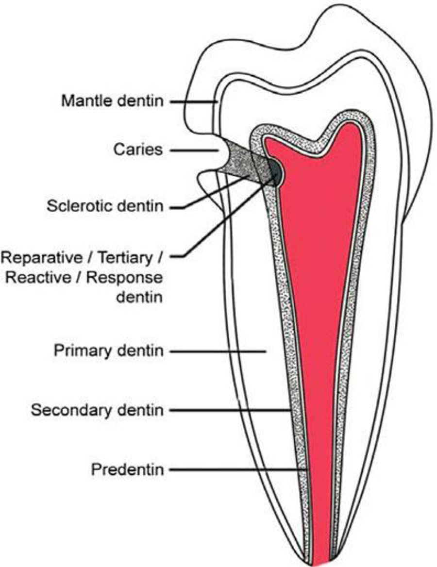
Dentin is classified into three types:
- Primary,
- Secondary, and
- Tertiary
PRIMARY DENTIN:
- Dentin formed before root completion
- Major part of dentin
- Mantle dentin – outer layer
- Circumpulpal dentin
| MANTLE DENTIN | CIRCUMPULPAL DENTIN |
| First formed dentin in the crown Seen just below the DEJ | Remaining portion of primary dentin which forms the bulk of the tooth |
| 20µm in thickness | Collagen fibrils are much smaller in diameter(0.05µm) |
| Organic matrix is formed by collagen fibrils arranged perpendicular to the DEJ | more mineralized than mantle dentin |
| Linear mechanism of calcification | Globular mechanism of calcification |
SECONDARY DENTIN
- Formed after root completion
- Narrow band of dentin bordering the pulp
- Contains fewer tubules than primary dentin
- There is usually a bend in the tubules where primary and secondary dentin interface
- Appears in greater amounts on the roof and floor of the pulp where it protects the pulp from exposure in older teeth
TERTIARY DENTIN
- Localized formation of Dentin At pulp –Dentin Border in response to noxious stimuli- Caries, Trauma Attrition, Cavity Preparation etc.
- It is of two types, either reactionary, where dentin is formed from a preexisting odontoblast, or reparative, where newly differentiated odontoblast-like cells are formed due to the death of the original odontoblasts, from a pulpal progenitor cell.
- Tertiary dentin is only formed by an odontoblast directly affected by a stimulus; therefore, the architecture and structure depend on the intensity and duration of the stimulus, e.g., if the stimulus is a carious lesion, there is extensive destruction of dentin and damage to the pulp, due to the differentiation of bacterial metabolites and toxins.
- Thus, tertiary dentin is deposited rapidly, with a sparse and irregular tubular pattern and some cellular inclusions; in this case, it is referred to as "osteodentin".
- Osteodentin can be found when there is a Vitamin A deficiency observed during development. When the stimulus is less present, nevertheless, it is laid down more slowly, with a normal tubular pattern and few cellular inclusions.


