- NEED HELP? CALL US NOW
- +919995411505
- [email protected]

Conventional tomography is a radiographic technique, usually using film, designed to image a slice or plane of tissue. This is accomplished by blurring the images of structures lying outside the plane of interest through the process of motion “ unsharpness. ”
Since CT, MRI, and cone-beam imaging, which have superior contrast resolution, film-based tomography has been used less frequently.
Uses in dentistry
When conventional tomography is used in dentistry it is applied primarily to high-contrast anatomy, such as that encountered in TMJ and dental implant imaging.
Mechanism of action
- Conventional tomography uses an x-ray tube and radiographic film rigidly connected and capable of moving about a fixed axis or fulcrum.
- The examination begins with the x-ray tube and film positioned on opposite sides of the fulcrum, which is located within the body’s plane of interest (focal plane).
- As the exposure begins, the tube and fi lm move in opposite directions simultaneously through a mechanical linkage.
- With this synchronous movement of tube and film, the images of objects located within the focal plane (at the fulcrum) remain in fixed positions on the radiographic film throughout the length of tube and fi lm travel and are clearly imaged.
- On the other hand, the images of objects located outside the focal plane have continuously changing positions on the film; as a result, the images of these objects are blurred beyond recognition by motion unsharpness.
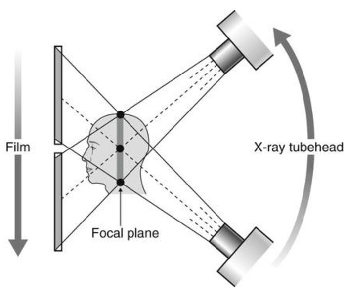
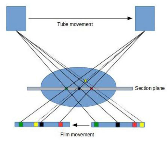
The resulting zone of sharp image is called the tomographic layer. Blurring of overlying structures is greatest (and the tomographic layer the thinnest) under the following circumstances:
- Overlying structures lie far from the focal plane.
- The focal plane lies far from the film.
- The long axis of the structure to be blurred is oriented perpendicular to the direction of tube travel.
- The distance of tube travel is large.
There are at least five types of tomographic movement:
- Linear,
- Circular,
- Elliptic,
- Hypocycloidal, and
- Spiral
Mechanically, the simplest tomographic motion is linear. More complex movements such as circular, elliptic, hypocycloidal, and spiral produce images without streaking artifacts common to the linear movements.
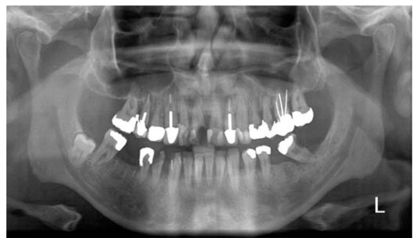
Many of the more expensive panoramic units are capable of making tomographic sections of the jaws.


