- NEED HELP? CALL US NOW
- +919995411505
- [email protected]
Formerly classified as Actinobacillus actinomycetemcomitans, this species was subsequently named Haemophilus actinomycetemcomitans, and is now known as Aggregatibacter actinomycetemcomitans. It is a major pathogen in juvenile
periodontitis.
A. actinomycetemcomitans (Aa) was first reported by Klinger in 1912 where he described a previously unknown Gram-negative microorganism that was found in actinomycotic lesions associated with Actinomyces, hence the latin word
“comitans” in common with Actinomyces.
It is a fastidious, non–spore-forming, nonmotile, facultatively anaerobic gramnegative rod that frequently complicates actinomycosis caused by Actinomyces israelii.
Often found in association with aggressive periodontitis, a severe infection of the periodontium.
Suspected to be involved in chronic periodontitis.
Less frequently, A. actinomycetemcomitans is associated with nonoral infections such as endocarditis.
| Its role in aggressive periodontitis was first discovered by Danish-born periodontist Jørgen Slots, a professor of dentistry and microbiology at the University of Southern California School of Dentistry |
Infection by this bacterium often is not resolved by the normal host, possibly because of inefficient phagocytosis and a weak oxidative burst response of neutrophils.
In addition, the bacterium has several virulence factors, including induction of apoptosis and tissue destruction.
Microbiology
- Actinomycetemcomitans grows slowly at 37degree C, aerobically or anaerobically, in standard broth media or on non-inhibitory solid media provided there is an atmosphere of approximately 5% carbon dioxide.
- In liquid media the organism tends to grow in small granules adhering to the walls of the bottle and this property is behind the name of the new genus Aggregatibacter .
- On agar, colonies generally become visible after 24 hours reaching approximately 3mm diameter after several days.
- Initially smooth, domed and translucent they become corrugated, starshaped, adherent and may pit the agar.
- Aggregatibacter actinomycetemcomitans is typically catalase positive and oxidase negative although occasional strains produce oxidase.
- It reduces nitrates to nitrites and is ONPG, urease and indole negative.
- It can ferment carbohydrates including glucose, fructose, maltose and mannose, and variably xylose, and mannitol.
- It is unable to ferment galactose, lactose, raffinose, sorbitol, sucrose, trehalose and glycerol
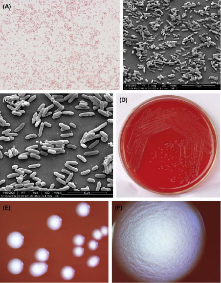
Figure
(A) Cells of A. actinomycetemcomitans (Gram stain).
(B) Cells of A. actinomycetemcomitans (SEM).
(C) Cells of A. actinomycetemcomitans (SEM). A. actinomycetemcomitans are 0.5–0.8 μm × 0.6–1.4 μm in size. Cells are spherical, club-shaped, or rod-shaped. Rod-shaped cells are common in agar cultures. The cells arrange as single cells, in pairs, or in piles. They produce no spores, are nonmotile, and do not form capsules. Cells stain gram-negative.
(D) A. actinomycetemcomitans colonies (BHI blood agar).
(E) A. actinomycetemcomitans colonies (stereomicroscope).
(F) A. actinomycetemcomitans colonies (stereomicroscope). On the surface of the agar, A. actinomycetemcomitans forms small colonies, with a diameter of approximately 0.5–1.0 mm. Primary cultures are often difficult to lift off the agar surface. Typical colonies are star-shaped or shaped like crossed cigars, with irregular edges. In broth culture, the growth shows small particle-like opacity and often sticks to the flask wall. However, some strains grow into a homogeneously turbid culture after repeated cultures.