- NEED HELP? CALL US NOW
- +919995411505
- [email protected]
DEVELOPMENT OF THE MANDIBLE

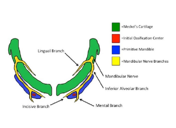
In the sixth week of IUL, Meckel’s cartilage extends as a solid rod of hyaline cartilage from the otic capsule to the midline.

- The right- and left-side cartilages are separated in the midline by a thin band of mesenchyme and the cartilage is surrounded by a fibrous capsule.
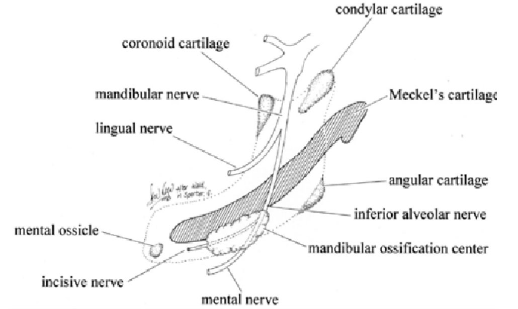
- The inferior alveolar and lingual branch of the mandibular nerve runs along the medial and lateral aspect of the cartilage.
- The inferior alveolar nerve bifurcates to form the incisive and mental branches at the site of the future mental foramen.
- Later, a mesenchymal condensation appears on either side near the bifurcation of the inferior alveolar nerve at the site of future mental foramen.
- In the seventh week of IUL, a single ossification centre for each half of the mandible begins in the mesenchymal condensation .
- The mandible is the second bone after the clavicle to ossify in the entire skeletal system.
- The ossification spreads around the inferior alveolar nerve towards the point of the future mandibular foramen posteriorly and towards the midline anteriorly.

- The ossification forms a trough around the inferior alveolar nerve and is later converted into the mandibular canal in which the nerve lies.
- From this bony canal, the alveolar plates develop in relation to the developing tooth germs.
- After the formation of the body of the mandible, the ramus is formed by the spread of ossification posteriorly into the mesenchyme of the first arch.
- The mandible formation is almost complete by the end of the 10th week, formed almost entirely by intramembranous ossification, and is entirely independent of Meckel’s cartilage.
- The union of the two separate centres of ossification occurs in the midline between the 4th and the 12th month postnatally.
- The dorsal end of Meckel’s cartilage ends in the tympanic cavity of the middle ear and is surrounded by the developing petrous part of the temporal bone.
| The auditory ossicles, incus and malleus, of the inner ear are derived from Meckel’s cartilage. |
- A part of the cartilage transforms to form the anterior malleolar ligament.
- During the 24th week of IUL, the major portion of Meckel’s cartilage between the mental foramen and mandibular foramen degenerates while the fibrocellular capsule of the cartilage persists between the lingula of the mandible and the spine of the sphenoid as the sphenomandibular ligament.
- Some remnants forming accessory endochondral ossicles are evident in the symphyseal region ventral to the mental foramen
- The growth of the mandible further takes place by the appearance of three secondary cartilages: condylar coronoid and symphyseal cartilages.
- These cartilages are developmentally and functionally different from Meckel’s cartilage, which is a primary cartilage The condyle appears as a cone-shaped cartilage during the 10th week of IUL, occupying most of the developing ramus
- The condylar head increases in size by both interstitial and appositional growth.
- The first evidence of endochondral ossification is seen in the 14th week of IUL.
- By the 20th week, most of the cartilage is replaced by bone and only a thin layer of cartilage remains in the head of the condyle.
- The remnant of the cartilage is present in adult life as a growth cartilage and articular cartilage of the temporomandibular joint.
- The coronoid cartilage appears in the 14th week of IUL, contributing to the development of the coronoid process.
- It soon becomes incorporated into the developing ramus and disappears before birth
- Two islands of symphyseal cartilage appear on either side of the midline in the symphyseal region independent of Meckel’s of IUL but as a variant some remnants may be present as mental ossicles in the fibrous union of the mandibular mid-line at birth.
- These ossicles ossify and are incorporated into the bone postnatally, during which the fibrous union (syndesmosis) between the two halves of the mandible is converted into a bony union (synostosis).
- Cartilage -It ossifies in the seventh month
Related posts
April 10, 2025
April 9, 2025
April 4, 2025


