- NEED HELP? CALL US NOW
- +919995411505
- [email protected]
Important Terminologies Of Enamel And Dentin
Dentinoenamel Junction
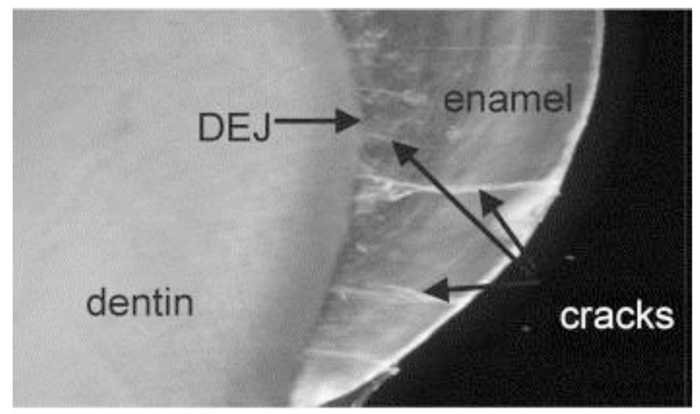
- The dentinoenamel junction (DEJ) is a scalloped border between the dentin and the enamel with the convexity of the scallop directed towards the dentin.
- Its scalloped nature increases the surface area of contact between the enamel and the dentin and reduces the incidence of cracks developing along the DEJ.
- The DEJ is less mineralized than the enamel and the dentin.
Enamel Lamellae
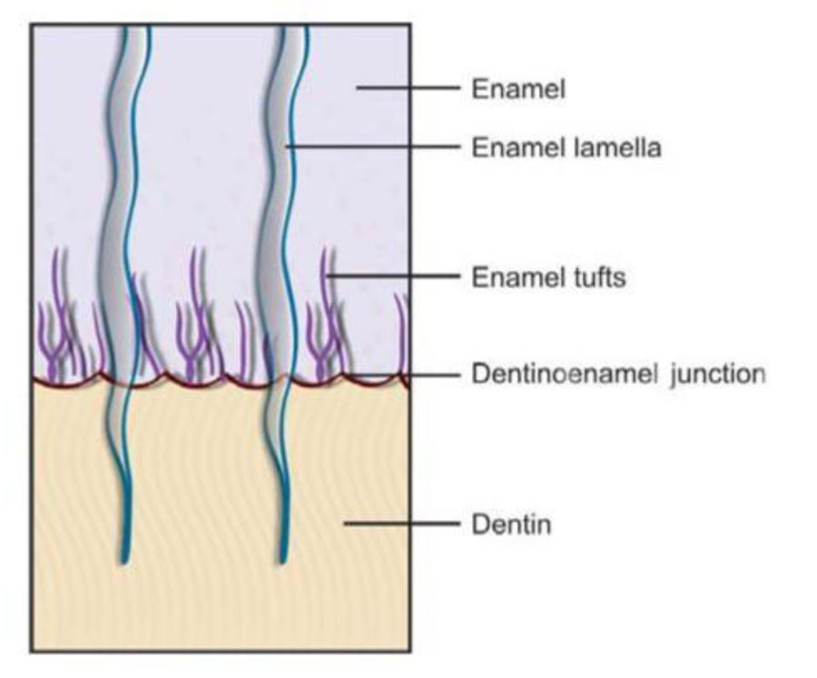
- Enamel lamellae are hypocalcified leaf-like structures extending from the outer surface of enamel towards the DEJ.
- These are formed due to stress occurring during enamel formation or due to cracks which develop in the enamel after eruption of teeth.
- These contain salivary components and organic debris.
- These are considered weak points in enamel and may allow passage of microorganisms which enter and initiate dental caries.
Enamel Tufts
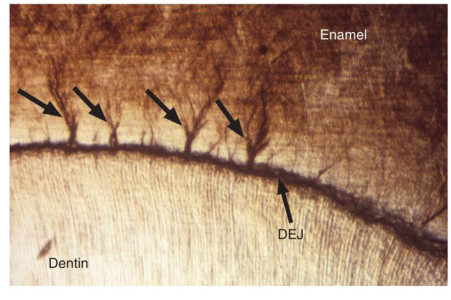
- Enamel tufts are hypocalcified structures appearing like tufts of grass at the DEJ.
- They extend up to one-thirds of the thickness of the enamel from the DEJ.
- These are areas in mature enamel where the highest protein content is present.
Enamel Spindles
- Enamel spindles are odontoblastic processes which pass across the DEJ into the enamel and become embedded in the enamel matrix.
- These appear as dark spindle-shaped structures due to thickening of their ends.
- These are more abundant in the cuspal region.
- These are easily identifiable in longitudinal section due to their alignment.
Incremental Lines of Retzius
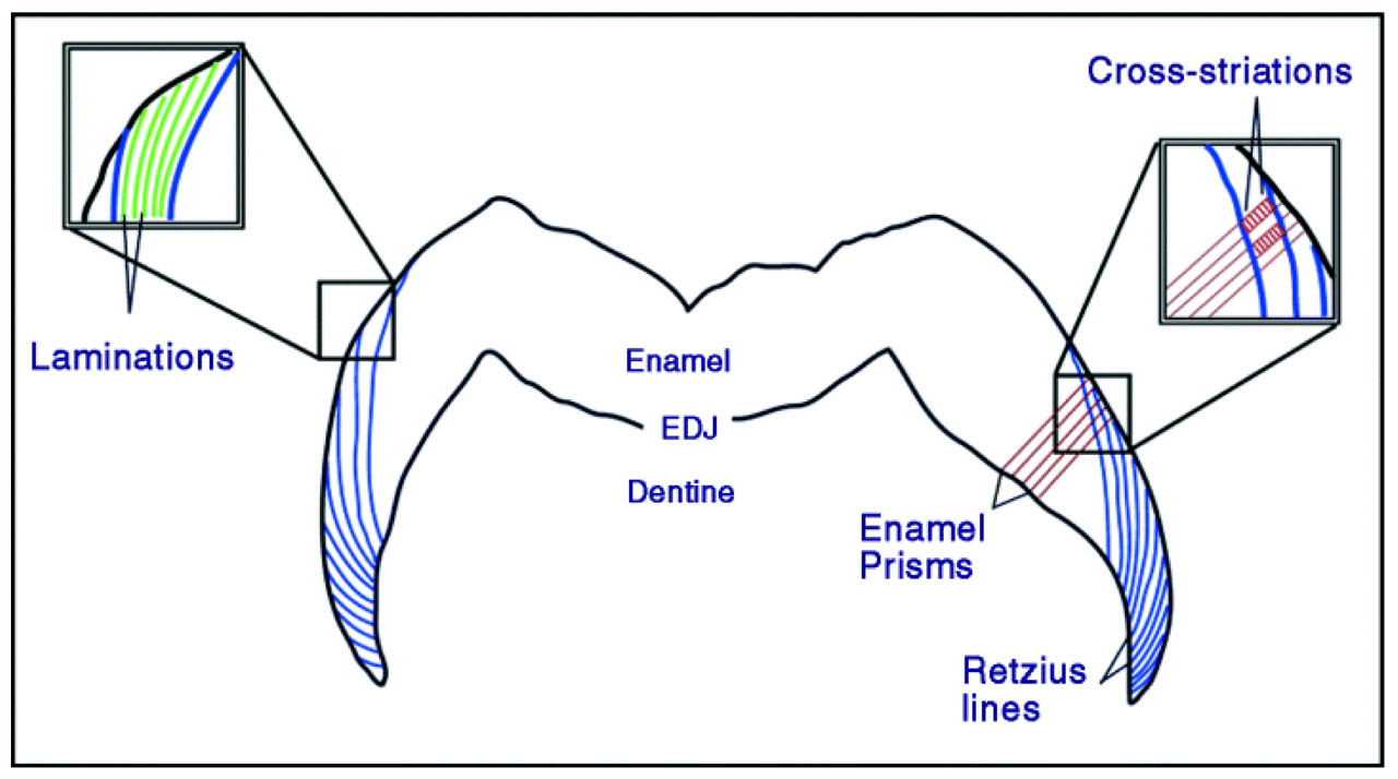
- The incremental lines of Retzius indicate the incremental or rhythmic deposition of enamel.
- These are seen as brownish lines that run obliquely across the enamel from DEJ to the outer surface of enamel.
- The striae are wider apart in the middle portion of the enamel, while they are closer together and more numerous in the cervical enamel due to the slower deposition of enamel.
- In cross-section, these appear as concentric rings.
- The lines are less prominent in the enamel formed before birth and more pronounced in the enamel formed after birth.
Dead Tracts
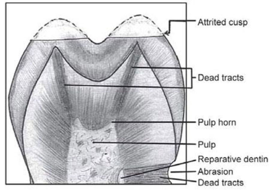
- Injury resulting in death and subsequent loss of odontoblastic process leads to empty dentinal tubules which become filled with air. Such tubules form dead tracts.
- In ground sections, these tubules appear dark in transmitted light and white in reflected light.
- Because of the convergence of the dentinal tubules towards the pulp, dead tracts appear narrow at the pulpal end and broader at the cuspal or incisal ends.
Interglobular Dentin
- Interglobular dentin is a zone of hypocalcification or hypomineralization seen in circumpulpal dentin in the crown and occasionally below the Tomes’ granular layer in root dentin.
- In ground sections, this appears as dark, star-shaped structure under transmitted light.
- This occurs due to the failure of globules of dentin to fuse into a homogenous mass during calcification.
- The dentinal tubules pass uninterrupted through interglobular dentin.
- Peritubular dentin is absent in those portions of the tubules passing through interglobular dentin.
Secondary Dentin
- Secondary dentin is formed after root completion.
- It is found bordering the pulp.
- It contains lesser number of tubules when compared with that of primary dentin.
- Tubules are more irregular when compared with that of primary dentin.
- Continuous deposition of secondary dentin on the floor of the pulp leads to smaller pulp chambers and narrower root canals.
Related posts
April 10, 2025
April 9, 2025
April 4, 2025


