- NEED HELP? CALL US NOW
- +919995411505
- [email protected]

- Obtaining autogenous, nonvascularized bone for the reconstruction of hard tissue defects and for the replacement of mandibular condyles.
- Obtaining a hyaline cartilage graft for the reconstruction of cartilaginous maxillofacial defects.
Indications of costochondral graft
- Temporomandibular joint (TMJ) replacements in pediatric patients with active growth centers to reconstruct condylar processes defects caused by trauma, neoplasms, infections, congenital dysplasias, growth abnormalities, ankyloses, and rheumatoid arthritis
- TMJ reconstruction in adult patients due to idiopathic condylar resorption, osteoarthritis, and rheumatoid arthritis when other methods (alloplastic joint replacement) are contraindicated
- Reconstruction of craniomaxillofacial defects caused by loss of hard tissue
- Reconstruction of skull defects or cranioplasty
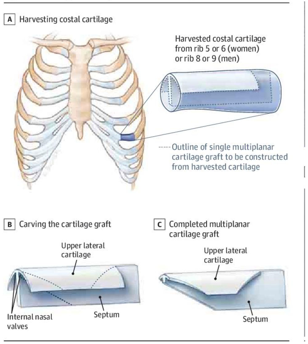
- Reconstruction of nasal dorsum defects or saddle nose deformities (costochondral cartilage)
- Reconstruction of the helical framework of the ear (costochondral cartilage)
Contraindications
- History of restrictive lung disease
- History of recent pulmonary infection
- History of cardiopulmonary instability
Advantages
- Biological compatibility
- Workability
- Functional adaptability
- Minimal additional detriment to the patient
- The growth potential of the costochondral graft makes it an ideal choice in children
Disadvantages
- Fracture
- Further ankylosis
- Increased operating time
- Additional surgical site
- Donor site morbidity
- Variable growth behavior of the graft
PROCEDURE
Costochondral Graft (CCG) Harvest Technique
- The anterior chest wall is prepped and draped, allowing for visualization of the sternum, clavicle, nipple, and umbilicus.
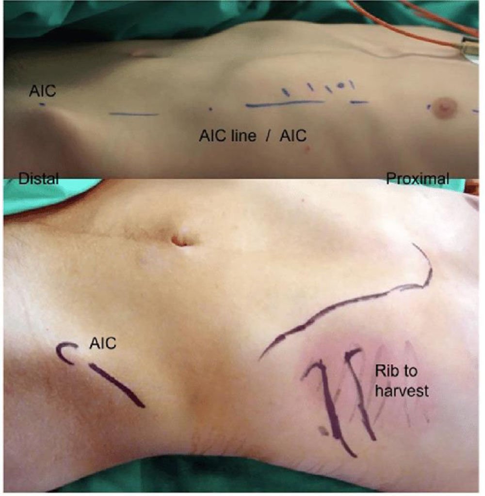
- The ribs are counted and marked with a marking pen.
A sterile marking pen is used to outline a 6–8 cm line within the infra mammary crease of female patients or at the level of the sixth or seventh rib in male patients. In pediatric female patients, the incision is placed in the anticipated future location of the inframammary crease.
A 6 cm incision is marked corresponding to the anticipated location of the inframammary crease, which corresponds to rib #6.
- Local anesthetic containing a vasoconstrictor is used to infiltrate the subcutaneous tissue overlying the rib to be harvested.
Digital pressure is used to identify the fifth, sixth, or seventh rib and the costochondral spaces. A 6–8 cm skin incision is made with a #15 blade directly over the superior aspect of the rib to be harvested. The incision transverses skin, subcutaneous tissue, and pectoralis muscle down to the perios- teum directly overlying the rib.
Dissection directly over the superior aspect of the rib to be harvested. The incision transverses skin, subcutaneous tissue, and pectoralis muscle.

A #9 periosteal elevator is used to dissect circumfer- entially around the rib. A tissue plane is developed between the rib’s periosteum–perichondrium and the thin parietal pleura . The subperiosteal dissection continues laterally as far as is needed and medially until the costochondral junction is reached. It is important to stay subperiosteal in order to avoid injury to the vascular bundle on the inferior portion of the rib.
A #9 periosteal elevator is used to dissect circumferen- tially around the rib. A tissue plane is developed between the rib's periosteum perichondrium and the thin parietal pleura.
- At the costochondral junction, dissection proceeds in a supraperichondrial plane so as not to detach the hyaline cartilaginous cap from the medial aspect of the rib.
- A guillotine rib cutter is used to transect the lateral portion of the rib.
Either a Doyen retractor or a silk suture is used to elevate the rib and to check the deep mar- gin for tissue–muscle adherence.
A silk suture is used to elevate the rib and to check the deep margin for tissue– muscle adherence after osteotomy of the lateral margin.
- A malleable retractor is placed deep to the medial aspect of the rib. A #10 blade is used to cut the medial aspect of the rib preserving a 5–10 mm cartilaginous cap. The malleable retractor will prevent the #10 blade from cutting into the underlying parietal pleura.
- The harvested costochondral graft is placed within a sterile, saline‐soaked gauze until the recipient site is prepared.

-
Once the rib or ribs are removed, sterile water is placed over the anterior chest wall defect, and the anesthesiologist is asked to provide positive pressure in order to inspect the harvest site for pleural perforations. If no air bubbles are present, then the harvest site is closed in layers. If minor air bubbles are present, pleural tears can be closed primarily with interrupted sutures. If large air bubbles are present, then a thoracotomy tube is placed.
Once the rib is removed, the anterior chest wall cavity is inspected for any bleeding or signs of pneumothorax. Sterile water can be placed over the anterior chest wall defect, and positive pressure is provided in order to inspect for pleural perforations.
- After closure of the surgical site in a layered fashion, steri-strips are applied to provide additional skin ten- sion. Generally, a drain is not required.
Complications
| Immediate or Early Complications | Late Complications |
Pleural tears, pneumothorax, and pleuritis Infection Hematoma or seroma formation Injury to the intercostal neurovascular Fracture at the bone–cartilage interface of the CCG |
Chest concavity: Occurs when multiple, adjacent ribs are harvested. Scar formation over the breast in female patients Areola retraction: Occurs when the incision is placed near the areolas. |


