- NEED HELP? CALL US NOW
- +919995411505
- [email protected]
Peripheral Eggshell Effect

Projection images, those that project a three-dimensional volume onto a twodimensional receptor, may produce a peripheral eggshell effect.
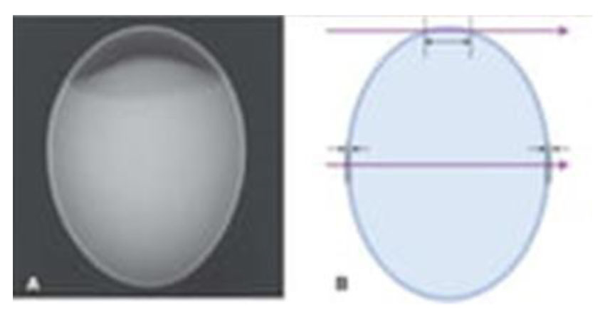
- The top photon has a tangential path through the apex of the egg and a much longer path through the shell of the egg than does the lower photon, which strikes the egg at right angles to the surface and travels through two thicknesses of the shell.
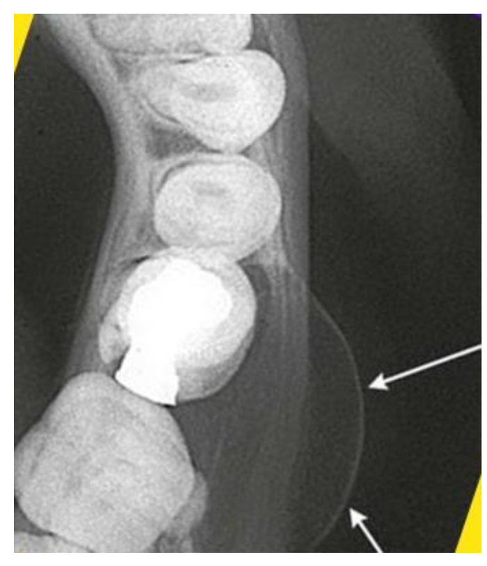
- As a result, photons traveling through the periphery of a curved surface are more attenuated, shows an expansile lesion on the buccal surface of the mandible on an occlusal view.
- Note how the periphery of the expanded cortex is more opaque than the region inside the expanded border.
- The cortical bone in not thicker on the cortex than over the rest of the lesion, but rather the x-ray beam is more attenuated in this region because of the longer path length of photons through the bony cortex on the periphery.
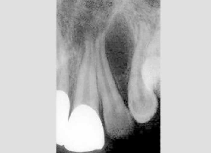
- This peripheral eggshell effect accounts for the lamina dura, the border of the maxillary sinuses and nasal fossa, and numerous other structures are well demonstrated on projection images.
- Note that soft tissue masses, such as the nose and tongue, do not show a peripheral eggshell effect because they are uniform rather then being composed of a dense layer surrounding a more lucent interior raveling at right angles to the surface.

