- NEED HELP? CALL US NOW
- +919995411505
- [email protected]
Anatomical Landmarks

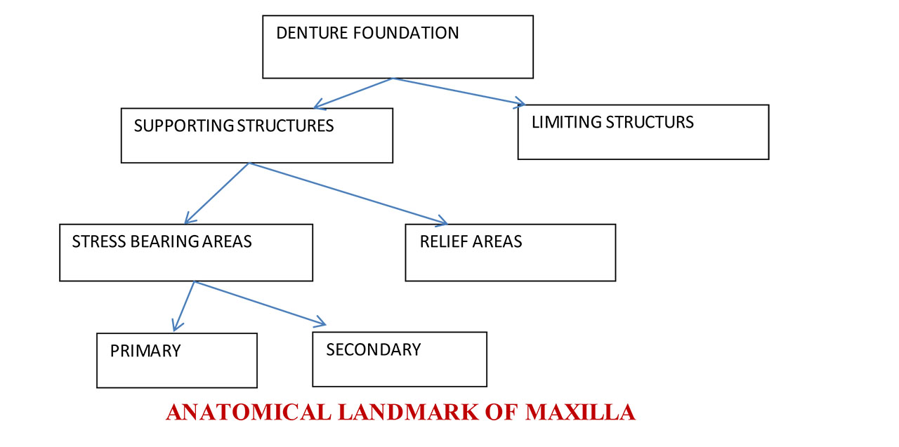
Supporting structures:
Primary stress bearing area / Supporting area:
- The horizontal portion of the hard palate lateral to the midline
- Posterolateral slopes and slopes of residual alveolar ridge
Secondary stress bearing area / Supporting area:
- Rugae area - set at an angle to residual ridge
- Maxillary Tuberosity
Limiting Structure Includes:
- Labial Frenum
- Labial Vestibule
- Buccal Frenum
- Buccal Vestibule
- Hamular Notch
- Posterior Palatal Seal Area
- Vibrating line
- Fovea palatinae
Relief Areas
- Incisive papilla
- Cuspid eminence
- Torus palatinus
- Zygomatic process
- Sharp spiny processes
- Mid-palatine raphe
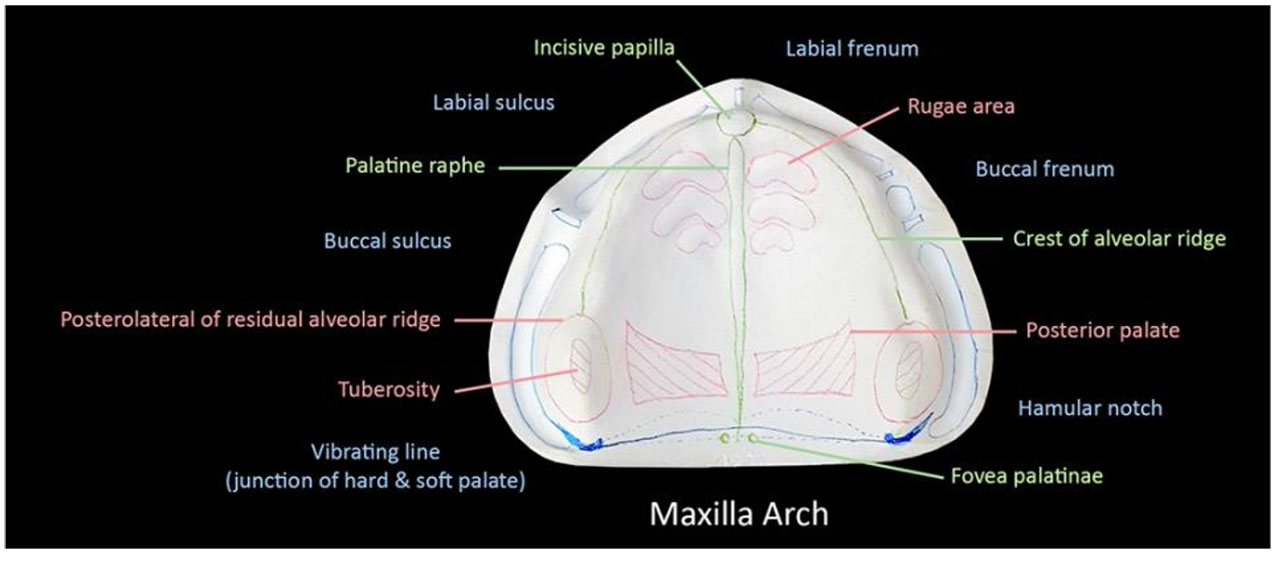
SUPPORTING STRUCTURES:
- Support is defined as ability to a denture to resist stresses that are directed towards the tissue
- These areas are the load-bearing areas. They show minimal ridge resorption even under constant load
- Denture should be designed such that most of the load is concentrated on these areas
- Hard palate
- Anterior region is formed by the palatine shelves of the maxillary bone
- Horizontal plate of the palatine bone forms the posterior part of the palate
- Submucosa in the mid-palatine suture is extremely thin
- Relief should be provided in the part of the denture covering the suture
- Horizontal portion of the hard palate lateral to midline acts as the primary support area
- Residual ridge
- The portion of the alveolar ridge and its soft tissue covering which remains following the removal of teeth
- Resorbs rapidly following extraction & continues throughout life in a reduced rate
- Sub-mucosa over the ridge has adequate resiliency to support the denture
- Crest of the ridge may act as secondary stress bearing area
- Posterolateral portion of the ridge is a primary stress bearing area
- Maxillary tubersity
- A bulbous extension of the residual ridge in the third molar region
- Posterior part of the residual ridge & tuberosity areas are most important areas of support because they are least likely to resorb
- Rugae
- Mucosal folds located in the anterior region of the palatal mucosa
- Acts as a secondary support area
- Folds of the mucosa play an important role in speech
- Hard palate
LIMITING STRUCTURES
They determine and confine the extent of the denture.
1. Labial Frenum
- It is a fold of mucous membrane extends from the mucosal lining of upper lip to the labial surface of the residual ridge.
- May be single or multiple, narrow or broad.
- Contains no muscle fibers and insert in a vertical direction which creates a maxillary labial notch in the maxillary impression or denture.
2. Labial vestibule
- It extends on both sides of the labial frenum to the buccal frenum, bounded by the upper lip and residual alveolar ridge.
- Contains no muscle fibers.
- In the denture the area that fills this space is known as labial flange.
3. Buccal Frenum
- A fold or folds of mucous membrane varies in size and shapes.
- It extends from the buccal mucous membrane reflection area toward the slope or crest of the residual alveolar ridge.
- Contains no muscle fibers and its direction antero-posteriorly.
- Produces the maxillary buccal notch in the maxillary impression or denture which must be broad enough because of the movement of the Frenum which is affected by some of the facial muscles as the orbicularis muscle pull it forward while buccinator muscle pull it backward.
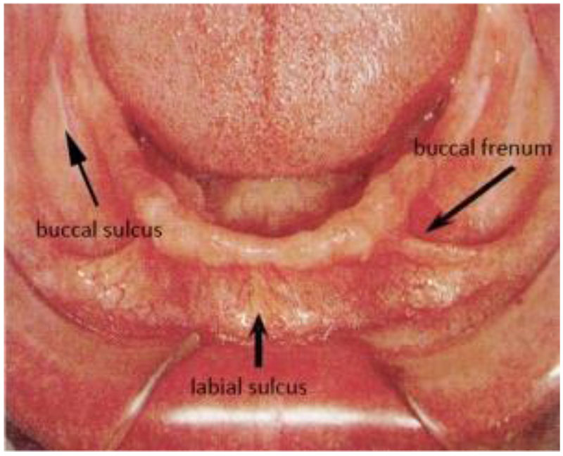
4. Buccal vestibule
- Is the space distal to the buccal frenum.
- Bounded laterally by the cheek and medially by the residual alveolar ridge.
- Area of the denture which will fill this space is known as buccal flange.
- Stability and retention of a denture are greater enhanced if the vestibule space properly filled with the flange distally.
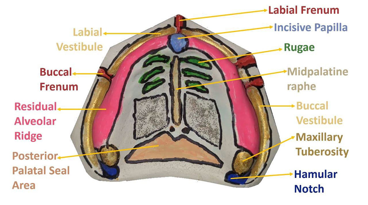
5. Hamular notch
- It is a narrow cleft of loose connective tissue situated between the maxillary tuberosity and the pterygoid hamulus (approximately 2mm antero-posteriorly).
- Used as boundary of the posterior border of maxillary denture.
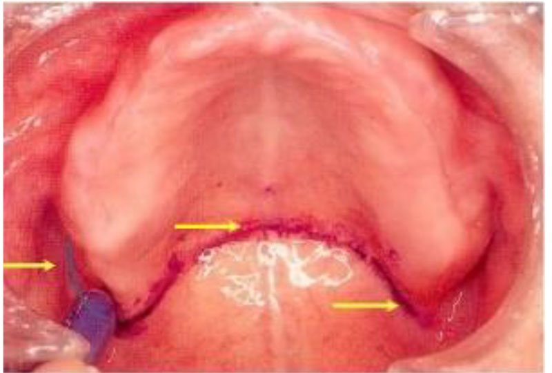
6.Vibrating line
- An Imaginary line drawn across the palate extended from one hamular notch tothe other.it is not well defined as a line
7. Posterior palatal seal area (post-dam)
- It is a soft tissue area at or beyond the junction of the hard and soft palates on which pressure within physiological limits can be applied by a complete denture to aid in its retention.
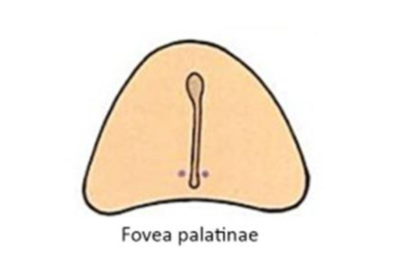
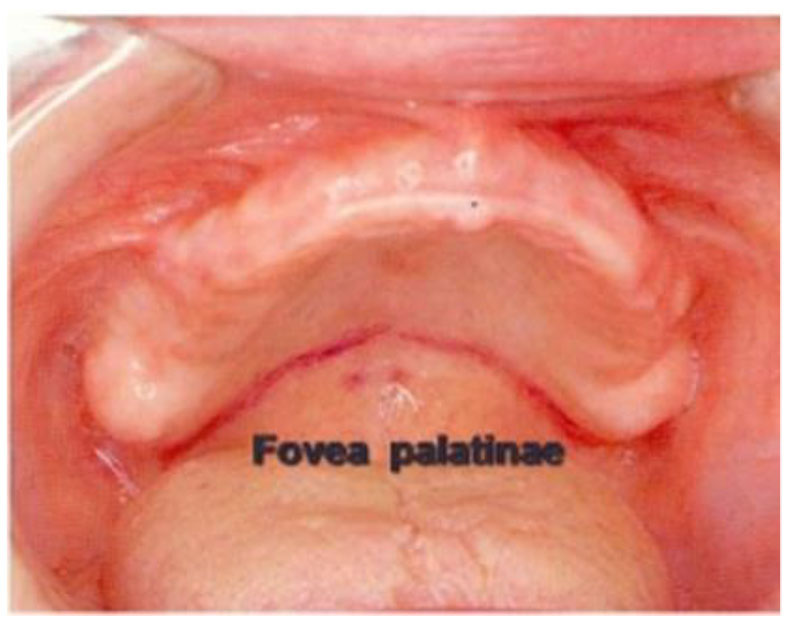
8. Fovea palatinae
- They are the remnants of ducts coalescence. Usually two in number on either side of the midline.
- They indicate the vicinity of posterior palatine seal area. It has no clinical significance
RELIEF AREAS
Denture should be designed such that the masticatory load is not concentrated over these areas
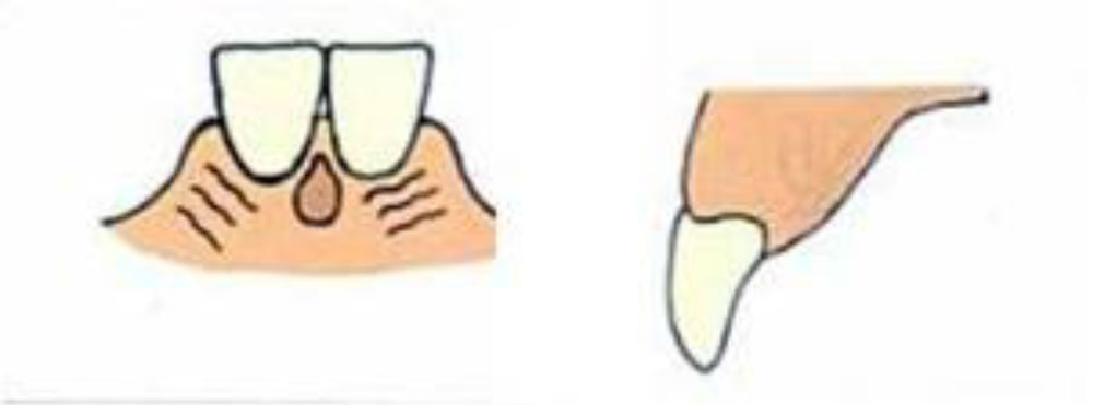
1. Incisive papilla
- A midline structure situated behind the maxillary central incisors
- It is a pad of fibrous connective tissue overlying the orifice of the nasopalatine canal.
- It is exit point of nasopalatine nerves & vessels
- Should be relieved
- Denture will compress the vessels or nerves & lead to necrosis of distributing areas & paraesthesia of anterior palate
2. Mid palatine raphe
- Median suture area covered by a thin submucosa
- Should be relieved during denture fabrication
- Most sensitive part of the palate to pressure
3. Sharp spiny spicules
- Spiny processes on maxillary and palatal bones deeply covered with soft tissue
- In resorbed ridge, these spicules irritate the soft tissue between them and denture base
- The canal leading from posterior palatine foramina often has sharp overhanging edge which may cut and irritate palatal soft tissue due to pressure from denture Sharp spiny spicules
4. Cuspid eminence
- Protruding bony ridge around maxillary canine
- Severe prominence must be provided relief
5. Zygomatic process
- It is close to residual alveolar ridge in malar region because of excess amount of resorption of alveolar ridge.
- It is thinly covered by mucous membrane.
- May cause soreness if not adequately relieved Zygomatic process Location: opposite first molar
Related posts
April 10, 2025
April 9, 2025
April 4, 2025


