- NEED HELP? CALL US NOW
- +919995411505
- [email protected]
Previous Years MCQ Discussion

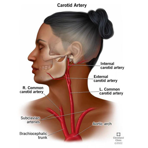
1. Common carotid artery is palpable at
- Upper border of thyroid cartilage
- Upper border of cricoid cartilage
- At hyoid bone
- Lower border of cricoid cartilage
ANSWER –a
- Carotid arteries are made up of three layers – the inner layer "tunica intima," the middle layer "tunica media," and the outer layer "tunica adventitia."
- The tunica intima consists of endothelium supported by a fragile elastic and also a collagenous layer of variable thickness.
- Smooth muscle comprises the tunica media, and it is responsible for changing the diameter of the blood vessel to regulate blood flow and blood pressure.
- The tunica adventitia attaches the carotid vessel to the surrounding tissue. The carotid arteries originate posterior to the sternoclavicular joints and in the neck, they are contained within the carotid sheath posterior to the sternocleidomastoid muscle.
- At the location of the upper border of the thyroid cartilage (typically at the level of the fourth or fifth cervical vertebra), the common carotid arteries bifurcate into the ECA and ICA.
- This bifurcation point is clinically significant as it serves as a point for the location of the "carotid body," a chemoreceptor, and the "carotid sinus," a baroreceptor.
- The carotid body chemoreceptor is sensitive to decreased PO2, increased PCO2, and decreased pH of blood, and is responsible for alerting the brain to change the respiratory rate. The carotid sinus baroreceptors respond to changes in the stretch of the blood vessel and are responsible for detecting changes and maintaining blood pressure
2. Most frequently damaged cranial nerve with a motor component
- Occulomotor
- V
- VII
- IX
ANSWER - c
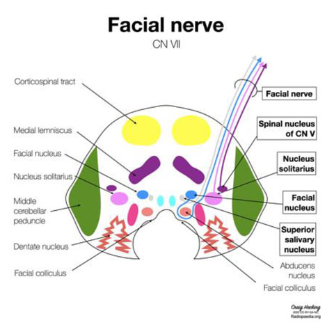
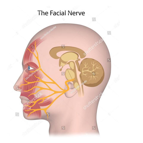
- The facial nerve is the seventh cranial nerve.
- It contains the motor, sensory, and parasympathetic (secretomotor) nerve fibers, which provide innervation to many areas of the head and neck region.
- The facial nerve is comprised of three nuclei :
- The main motor nucleus
- The parasympathetic nuclei
- The sensory nucleus
- There are four major functions of the facial nerve :
- General somatic efferent (motor supply to facial muscles)
- General visceral efferent (parasympathetic secretomotor supply to submandibular and sublingual salivary glands and the lacrimal gland)
- Special visceral afferent (taste sensation from anterior two-thirds of the tongue)
- General somatic afferent (cutaneous sensations from the pinna and the external auditory meatus).
- Motor Pathway
- The upper motor neuron is located in the facial motor area of the precentral gyrus.
- The axons from the upper motor neuron travel along the ipsilateral corticobulbar tract to the lower pons, where most fibers cross to the other side and synapse with the lower motor neuron.
- The main motor nucleus (lower motor neuron) divides into four subnuclei; dorsal, intermediate, lateral, and medial.
- In an upper motor neuron facial palsy, only the contralateral lower quadrant of the face is paralyzed, while the ipsilateral half of the face suffers paralysis in a lower motor neuron facial palsy.
- The main motor nucleus is responsible for the voluntary control of facial muscles.
- Motor nucleus supplies
- Auricular muscles,
- Posterior belly of the digastric muscle
- Stapedius muscle, and the stylohyoid muscle
3. Third nerve palsy causes all except
- Ptosis
- Miosis
- Outward eye movement
- Diplopia
ANSWER-b
- The third cranial nerve controls the movement of four of the six eye muscles. These muscles move the eye inward, up and down, and they control torsion (rotating the eye downward and toward the ear on the same side).
- The occulomotor nerve also controls constriction of the pupil, the position of the upper eyelid, and the ability of the eye to focus.
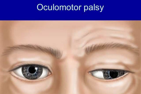
- A complete nerve palsy causes a completely closed eyelid and deviation of the eye outward and downward. The eye cannot move inward or up, and the pupil is typically enlarged and does not react normally to light.
- A partial third nerve palsy affects, to varying degrees, any of the functions controlled by the third cranial nerve.
- Symptom
- An enlarged pupil that does not react normally to light.
- Double vision (diplopia)
- Droopy eyelid (ptosis)
- Eye misalignment (strabismus)
- Tilted head to compensate for binocular vision difficulties.
4. Propioceptive fibres are not carried by which cranial nerve
- Cranial accessory
- Glossopharyngeal
- Trigeminal
- Facial
ANSWER-a
5. Intrinsic muscles of eyeball are all except
- Levator palpebrae superioris
- Ciliaris
- Sphincter pupillae
- Dilator pupillae
ANSWER-a
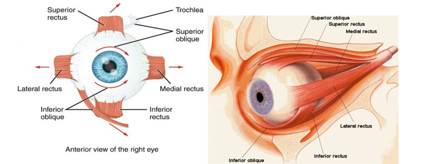
- There are two types of eye muscles: extrinsic muscles that control eye movement and position, and intrinsic muscles
Extrinsic eye muscles
- Are attached to the outside of the eyeball and enable the eyes to move in all directions of sight.
- There are six extraocular eye muscles and one muscle that controls movement in the upper eyelid.
- Superior rectus,
- Inferior rectus,
- Lateral rectus,
- Medial rectus,
- Superior oblique and Inferior oblique
- One muscle that controls eyelid elevation (levator palpebrae)
Intrinsic muscle
- That control near focusing and how much light enters the eye.they are
- Ciliary,
- Dilatator pupillae
- Sphincter pupillae muscles.
Related posts
April 10, 2025
April 9, 2025
April 4, 2025


